
Urolithin A: PINK1/Parkin mitophagy, muscle function, and limits of the evidence
From the microbial ellagitannin cascade to ubiquitin-mediated mitophagy and human trials: primary outcomes, secondary signals, pharmacokinetics, and urolithin A safety.
Urolithin A is often described with a single arrow: dietary ellagitannins → mitophagy → stronger muscle. Different biological systems and measurement methods lie between those steps. Bacterial transformation takes place in the colonic lumen, conjugation in enterocytes and the liver, recognition of damaged mitochondria inside cells, and muscle strength and walking are human outcomes. A coherent hypothesis does not mean that every link has been confirmed for an oral product in human muscle. This review examines the molecular mechanism in detail, then compares it with measured clinical outcomes and the limits of their interpretation.
1. From ellagitannin to a urolithin metabotype
1.1. Chemical starting material and its barriers
Ellagitannins are hydrolysable esters of hexahydroxydiphenic acid and sugars. They occur in pomegranate, some nuts, and berries, with amounts and composition depending on species, cultivar, and processing. Pomegranate punicalagin (C₄₈H₂₈O₃₀; approximately 1084.7 g/mol) illustrates why a large polar molecule is not equivalent to a small intestinal metabolite: absorption of the parent tannin is limited. Hydrolysis and lactonization release ellagic acid (C₁₄H₆O₈; approximately 302.2 g/mol). Its smaller size does not eliminate low aqueous solubility and variable absorption. Therefore, concentrations of the tannin itself, ellagic acid, free urolithin, and its conjugates cannot be substituted for one another when comparing food with a supplement.
The structure of urolithin A is 3,8-dihydroxy-6H-benzo[c]chromen-6-one (C₁₃H₈O₄; approximately 228.2 g/mol). It is a metabolite of the bacterial community, not an unchanged constituent of pomegranate juice. Food also carries other phenolic compounds, fibre, and nutrients, so differences in outcomes between a diet and isolated urolithin cannot be attributed entirely to the proportion of people who produce urolithin A.
1.2. Sequence of reactions in the gut
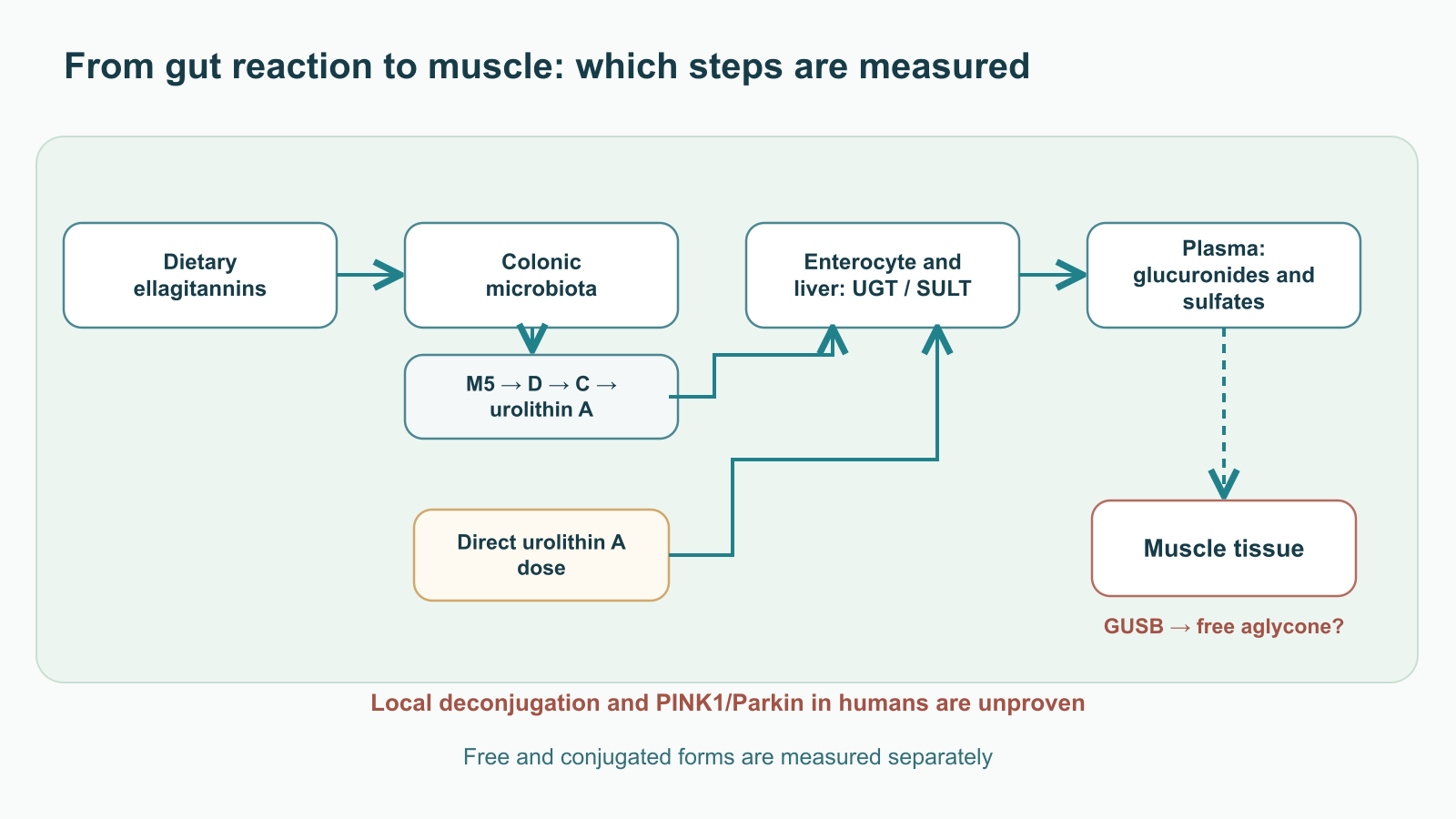
After ellagitannin hydrolysis, ellagic acid reaches intestinal regions where microbial lactone-ring opening, decarboxylation, and sequential dehydroxylation reactions generate a family of urolithins. The conversion map can be written schematically as follows:
This notation shows the chemical logic of losing hydroxyl groups, but it does not claim that every strain performs every step or that intermediates always appear in this sequence in every person. M5 is a pentahydroxylated intermediate; D and C have four and three hydroxyl groups, respectively; A and isourolithin A are positional dihydroxylated isomers. A further branch can lead to urolithin B. Hydroxyl positions affect interactions with enzymes and measured activity; the isomers cannot be declared equivalent.
The isolates Gordonibacter urolithinfaciens and Gordonibacter pamelaeae can carry out early stages of ellagic-acid transformation, while Ellagibacter isourolithinifaciens has been described as an isourolithin A producer. Detecting these taxa alone does not prove complete urolithin A synthesis: the product depends on the community’s functional composition, culture conditions, substrate, and transit time. The specific organism responsible for late dehydroxylation in humans cannot be reduced to one reliably established “obligate” species. Syntrophy is a useful experimental hypothesis, but attributing a step to uncultivated clostridia and a molybdenum dehydroxylase as a proven universal pathway would go beyond the available evidence.
1.3. Metabotypes A, B, and 0: phenotype, not diagnosis
After a standardized ellagitannin challenge, researchers distinguish UM-A, with predominant urolithin A production; UM-B, with isourolithin A and/or urolithin B alongside A; and UM-0, with no detectable urolithins at the analytical threshold used. Metabotype designation depends on diet before testing, sampling time, matrix (urine or plasma), assay sensitivity, and population. It should not be turned into an immutable genomic trait, a diagnosis of “dysbiosis,” or an indication for supplementation. Microbial associations, age, and metabolic disease may affect group distribution, but exact proportions of “non-producers” cannot be transferred across populations without using the same test.
| Metabotype after challenge | Analytical description | What can and cannot be concluded |
|---|---|---|
| UM-A | Urolithin A and its conjugates predominate | The microbial pathway is active under the test conditions; this does not prove lower inflammation or better muscle function. |
| UM-B | Isourolithin A and/or urolithin B appear alongside other metabolites | This is a different dehydroxylation profile; it cannot automatically be called “toxic” or incapable of producing A. |
| UM-0 | Urolithins are not detected | Microbiome features, exposure, or the analytical window may contribute; a zero result is not a proven clinical need for exogenous A. |
Direct intake of purified urolithin A reduces dependence on the microbial step, but does not make exposure identical across people: absorption, conjugation, transport, distribution, and elimination remain variable. A choice between food and a supplement cannot be inferred from metabotype without a trial linking that classification to a functional outcome.
2. How the cell recognizes a mitochondrion for removal
2.1. Inner-membrane potential and control of PINK1 import
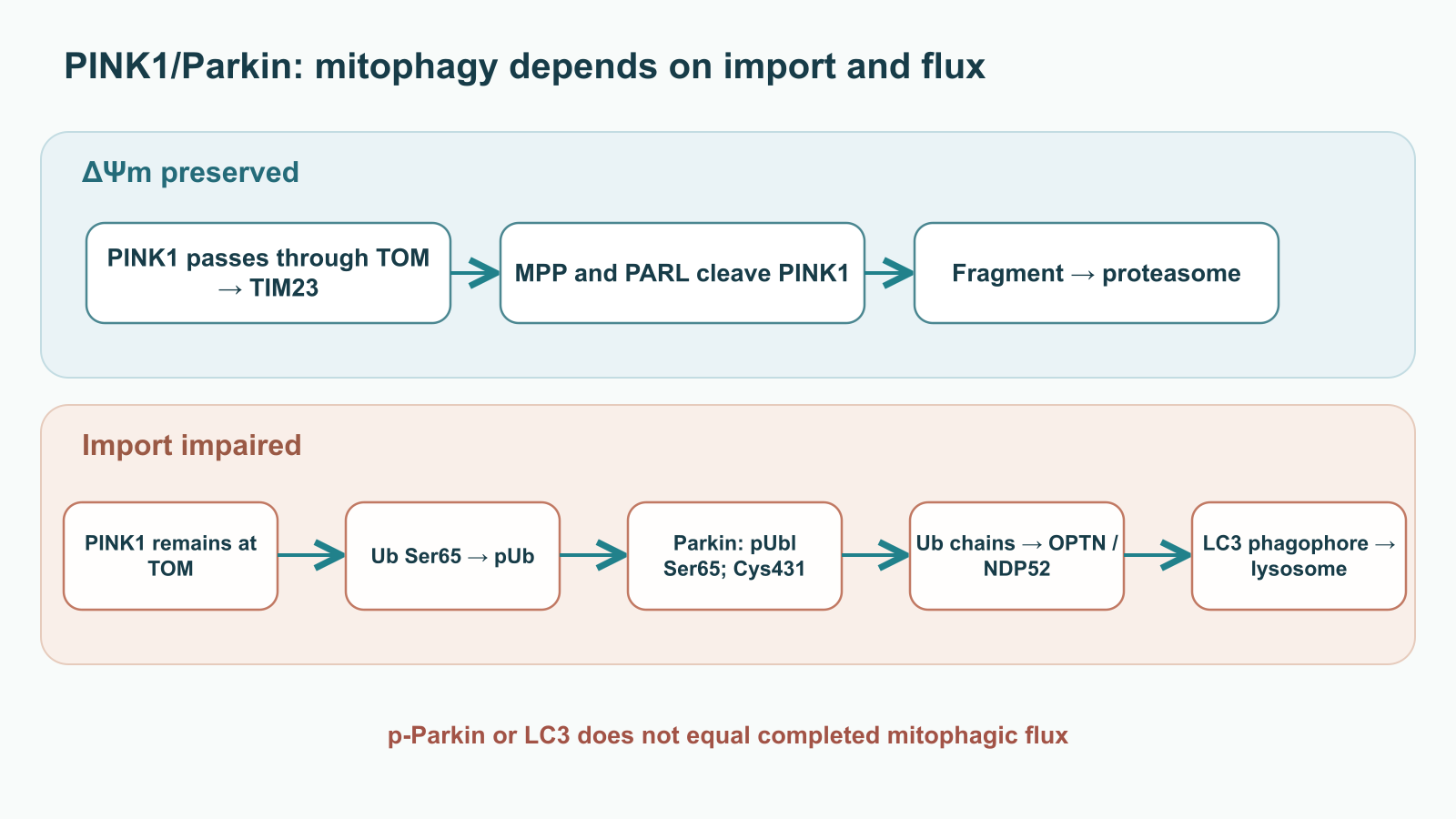
The mitochondrial membrane maintains an electrochemical proton gradient. Its electrical component, , helps import proteins with a positively charged N-terminal targeting sequence. PINK1, a serine/threonine kinase, passes through the outer-membrane translocase TOM and, when the potential is intact, engages the inner-membrane TIM23 system. Mitochondrial processing peptidase (MPP) removes the targeting segment; the protease PARL cleaves PINK1 in the inner membrane. The resulting fragment can return to the cytosol and undergo proteasomal degradation after recognition of its N-terminal phenylalanine. Consequently, little full-length PINK1 is found on the surface of a functioning, polarized organelle. “Little” does not mean absolute zero, and depolarization is not the only known form of mitochondrial damage.
When potential is lost sufficiently, import through TIM23 and access to PARL are disrupted. Full-length PINK1 is retained on the outer membrane in association with TOM and can phosphorylate accessible substrates from the cytosolic side. Experimental systems have described stabilization of the complex and trans-autophosphorylation of the kinase, including at Ser228 and Thr257. However, the precise stoichiometry of TOM subunits, complex mass, and a single threshold for depend on the model; they should not be turned into universal values for aging human muscle. Individual mitochondria vary in potential, shape, location, and degree of exchange with the network.
2.2. Phospho-ubiquitin Ser65 as a local tag
Basal ubiquitin tags are already present on outer mitochondrial membrane proteins. PINK1 transfers an ATP-derived phosphate to ubiquitin Ser65:
Phospho-ubiquitin, or pUb, changes the local binding surface and serves as a signal for Parkin recruitment. Structural studies show conformational shifts in ubiquitin and its binding domains; they do not justify treating pUb as resistant to every deubiquitinase. The ratio of phosphorylated to total ubiquitin depends on PINK1 kinase rate, phosphatase activity, chain extension, and chain removal. The local density of pUb on the membrane matters more for positive feedback than a single measurement of total cellular ubiquitin.
2.3. Ubl, REP, RING0–2: how Parkin is released
Parkin (PRKN) belongs to the RBR ligase family. Its N-terminal Ubl domain, RING0, RING1, IBR, RING2, and inhibitory REP element hold the enzyme in an autoinhibited conformation. RING1 participates in E2 binding, RING2 carries catalytic Cys431, and Ubl/REP contacts restrict access to these sites. pUb on the membrane binds Parkin and promotes rearrangement of interfaces; after Ubl is released, PINK1 phosphorylates its Ser65. Phosphorylated Ubl further relieves autoinhibition, allowing E2 to transfer ubiquitin first to Parkin Cys431 and then to the amino group of a substrate lysine. This thioester intermediate distinguishes the RBR mechanism from the simplified scheme in which “E2 transfers Ub directly to a membrane protein.”
Structural studies reveal the positions of REP and Ubl, but domain rotation angles and interatomic distances describe particular constructs and crystallization conditions. They cannot be transferred without qualification to the dynamics of a living cell. Parkin phosphorylation and pUb binding reinforce one another but are experimentally distinguishable stages. Even detecting p-Parkin in a biopsy does not prove completed lysosomal mitophagic flux.
2.4. Membrane substrates, different chain linkages, and receptors
Parkin modifies many outer-membrane proteins, including mitofusins MFN1/2, transport GTPases MIRO/RHOT, and VDAC. Extraction by p97/VCP and proteasomal degradation matter for MFN and MIRO: they can limit renewed fusion or movement of a damaged organelle. These are functional tendencies and do not always amount to shutting down the mitochondrial network. Ubiquitin chains include K6, K11, K48, and K63 linkages, which participate in different stages of signaling; the rule “K48 = proteasome only, K63 = autophagy only” would be too binary.
New membrane chains provide PINK1 with new Ub molecules to phosphorylate. An amplification loop emerges: pUb recruits Parkin, Parkin adds Ub, and PINK1 phosphorylates the added Ub. Deubiquitinases and phosphatases limit the loop, so damage does not have to trigger irreversible removal. Receptors OPTN, NDP52/CALCOCO2, TAX1BP1, and other proteins assemble on the modified surface; p62/SQSTM1 may participate, but its contribution is not obligatory in every model. OPTN and NDP52 recognize chains through ubiquitin-binding modules and connect them to the autophagy machinery. The kinase TBK1 phosphorylates OPTN and supports local recruitment, although the precise increase in affinity cannot be treated as fixed across all substrates and tissues.
2.5. Phagophore, LC3, and lysosome: flux matters more than the tag
A receptor connects the ubiquitinated organelle to the growing phagophore membrane by interacting with ATG8 proteins, including lipidated LC3. The general LIR-motif notation describes one class of contacts; the context of flanking amino acids and their phosphorylation changes affinity. Phagophore initiation and expansion require the ULK1 complex, PI3KC3, WIPI, and ATG-conjugation systems; the membrane cannot be attributed to a single, invariant source. After the mitophagosome closes, its fusion with the lysosome is aided by Rab7, SNARE proteins including STX17, SNAP29, and VAMP8, and other positioning proteins. V-ATPase creates an acidic environment in which cathepsins and nucleases break down the contents.
The number of LC3-positive structures or p-Parkin is a snapshot of an intermediate state. An increase in structures can reflect either greater delivery or a block in their clearance. To infer mitophagic flux, measurements must account for the lysosomal stage, for example with dynamic reporters or comparison under degradation blockade in an appropriate experimental model. The PINK1/Parkin pathway is well characterized, but Parkin-independent receptor-mediated mitophagy mechanisms also exist. Urolithin A can change related markers in preclinical models; this does not establish that the entire sequence above is a necessary and proven mechanism for every clinical effect.
3. What happens after an organelle is removed
3.1. AMPK, SIRT1, and PGC-1α: coordination, not automatic replacement
Mitochondrial removal has physiological value only when sufficient energy-producing mass and network structure are maintained. Cellular energy stress is reflected in the AMP/ATP and ADP/ATP ratios. AMP and ADP bind the regulatory γ-subunit of AMPK; phosphorylation of Thr172 in the catalytic α-subunit, including by LKB1, increases complex activity. AMPK changes substrate use and can phosphorylate the coactivator PGC-1α. This is general regulatory physiology, not an established sequence in which “urolithin A always lowers ATP first and then renews all mitochondria.” Such an ATP decrease may also be harmful; AMPK activity itself depends on tissue, nutrition, workload, and observation time.
SIRT1 uses NAD⁺ as a deacetylation substrate, including for PGC-1α. NAMPT participates in nicotinamide salvage; the NAD⁺/NADH ratio, compartments, and NAD⁺ consumption differ among the cytosol, nucleus, and matrix. AMPK–PGC-1α–SIRT1 is best considered a connected network rather than one obligatory linear reaction. PGC-1α coactivates NRF1, ERR and PPAR receptors; nuclear programs increase expression of respiratory components, protein-import machinery, and TFAM. Import of TFAM into the matrix supports mtDNA packaging and transcription. Biogenesis also includes lipid synthesis, protein import, complex assembly, mtDNA replication, and network remodeling: a change in one transcriptional signature does not measure the total number of new functional organelles.
In muscle, exercise itself changes AMPK, PGC-1α, the mitochondrial network, and capillaries. Attributing an effect to a supplement acting through this axis requires an intervention, a time course, and functional assessment—not merely two markers changing together after a course. Urolithin A trials measured selected protein and transcriptomic signatures; these should be read as signs of a possible tissue response, not direct observation of the complete sequence mitophagy → biogenesis → recovery of strength.
3.2. How mtDNA may activate cGAS–STING
When membranes are damaged or quality control is defective, mtDNA may enter the cytosol. The permeability transition pore (mPTP) in the inner membrane, outer-membrane disruption, BAX/BAK-dependent macropores, and other processes may contribute to mtDNA release under different conditions; there is no single obligatory channel. Cytosolic DNA binds cGAS, which produces 2′3′-cGAMP from ATP and GTP. This cyclic dinucleotide activates STING on the endoplasmic reticulum. STING trafficking toward the Golgi and recruitment of TBK1/IRF3 lead to transcription of interferon genes; NF-κB signaling adds inflammatory components. Bacterial-like CpG motifs in mtDNA do not replace the key condition: the DNA must reach a compartment accessible to the sensor.
Animal and cell models show that PINK1/Parkin can limit STING-dependent inflammation during mitochondrial stress. However, available clinical CRP and cytokine measurements cannot support the claim that oral urolithin A “blocks” this axis in an older person. CRP is a nonspecific systemic marker; it does not localize mtDNA release, cGAS activity, or the source of cytokines. The causal chain can be clarified only by measuring relevant intermediate events in an appropriate model and, separately, in human tissue.
3.3. NLRP3, pyroptosis, and the limits of the term inflammaging
A second possible link between defective mitochondria and inflammation is the NLRP3 inflammasome. Its activation is usefully divided into priming and activation. NF-κB-dependent priming increases expression of NLRP3 and pro-IL-1β; subsequent ionic, metabolic, and organellar signals support assembly of NLRP3 with the ASC adaptor and procaspase-1. Active caspase-1 processes pro-IL-1β and pro-IL-18, while gasdermin D cleavage creates pores associated with pyroptosis. Oxidized mtDNA and mitochondrial ROS are considered possible participants in activation, but a universal direct binding of ox-mtDNA to a specific NLRP3 domain and an obligatory heptameric scaffold should not be asserted for every tissue.
The senescence-associated secretory phenotype, SASP, includes more factors and contexts than a single inflammasome. In muscle, inflammatory mediators can affect IGF-1/Akt/mTOR, proteolysis, MuRF1 and atrogin-1 activity, and the stem-cell niche. Sarcopenia also arises from other causes: denervation, reduced activity, disease, inadequate nutrition, hormonal and vascular changes. Therefore, even if some inflammatory markers decrease, one cannot conclude that a treatment has eliminated “inflammaging,” restored myosin, or prevented sarcopenia.
4. Muscle as a system: different compartments and different outcomes
4.1. Myocyte mitochondria and multifactorial sarcopenia
Subsarcolemmal mitochondria lie closer to the sarcolemma and signaling at its surface; intermyofibrillar mitochondria are distributed among contractile elements, supporting local ATP and calcium exchange. The distinction between mitochondrial ATP production and the brief phosphocreatine buffer helps explain why an endurance response cannot be substituted for a maximal-strength response; the buffer mechanism is described in the article on creatine and muscle performance. These are functional compartments, but they are interconnected. Fusion and fission dynamics, the respiratory chain, cristae content, fatty-acid transport, and quality control affect the muscle’s ability to sustain workload. Incomplete β-oxidation and changes in acylcarnitines may signal metabolic remodeling; plasma acylcarnitine concentration is not a direct measure of ATP in a myofibril. Diacylglycerols, ceramides, and PKCθ participate in lipotoxic disruption of insulin signaling, but a causal link between each and a specific urolithin A dose requires separate testing.
Sarcopenia is assessed primarily through muscle strength, mass, and physical function using criteria for a specific population. Mitochondrial dysfunction may contribute to its development but is not its sole diagnostic mechanism. Urolithin A trials included people with different baseline characteristics; strength data from healthy or sedentary volunteers cannot automatically be transferred to patients with diagnosed sarcopenia. Six-minute walk performance also depends on the heart, lungs, joints, motivation, and practice effects from repeat testing.
4.2. Satellite cells and conditions for regeneration
Pax7⁺ satellite cells lie between a muscle fibre’s sarcolemma and its basal lamina. At rest they retain self-renewal capacity; after injury, some activate Myf5/MyoD, proliferate, and differentiate with the participation of myogenin, while others return to the reserve pool. Mitochondrial dynamics, autophagy, and ROS participate in transitions between these states. Elevated ROS should not always be considered pathological: a brief signal is needed for regeneration, whereas chronic damage and impaired quality control may limit it. p16INK4a and p21CIP1 are markers of context-dependent senescence and cell-cycle-arrest programs, not direct measures of muscle strength.
Preclinical models support the hypothesis that improved mitochondrial quality control in Pax7⁺ cells may help preserve the regenerative response. However, clinical trials of urolithin A do not demonstrate restoration of the human MuSC pool or formation of new mature myotubes in patients. Such a conclusion would require biopsies with cellular markers, lineage tracing, and functional assessment of regeneration, ideally after standardized injury and with training controlled.
4.3. Type I, IIa, and IIx fibres, perfusion, and endurance
Type I fibres on average have high oxidative capacity and fatigue resistance; type IIa fibres combine fast contraction with a substantial aerobic component; type IIx fibres rely more strongly on glycolysis. Fibre-type distribution depends on muscle, age, innervation, and training. Treating human type IIb fibres as a separate universal group would be inaccurate. Aging especially affects fast motor units, but the clinical phenotype cannot be inferred from a single “type shift.”
PGC-1α-related programs may affect CPT1B, CD36, oxidation enzymes, and VEGF-A. This connects mitochondrial metabolism with capillarization as a hypothesis and in experimental models. CPT1B regulates entry of long-chain acyl groups through the carnitine shuttle, while CD36 mediates fatty-acid uptake; the constraint at this step imposed by malonyl-CoA is discussed in more detail in the article on CPT1 and β-oxidation. A lower post-exercise lactate level depends not only on pyruvate dehydrogenase but also on work intensity, tissue metabolism, and lactate clearance. Urolithin A trials have not shown a reliable increase in the capillary-to-fibre ratio, obligatory conversion of IIx to I/IIa, or proven acceleration of lactate clearance. Endurance should be assessed using a prespecified test and between-group effect, not a plausible fibre diagram.
5. What human intervention studies have shown
Clinical trials answer questions about a particular measured outcome at a particular dose and duration. A change in acylcarnitines, mitophagy-related protein expression, or post-exercise well-being does not replace a prespecified functional endpoint. Several randomized studies are available in this field, but sample sizes are small and long-term trials in patients with diagnosed sarcopenia are insufficient. The product and some studies are linked to a commercial sponsor; this calls for especially clear separation of primary and secondary results, not automatic dismissal of the data.
5.1. Andreux 2019: tolerability and molecular signatures
The first human study enrolled 60 healthy, sedentary older volunteers. A single- and repeated-dose protocol assessed safety, pharmacokinetics, and molecular measures during short exposure; repeated dosing lasted 28 days. Participants received oral urolithin A at studied doses that included 250, 500, and 1000 mg/day. Analysis of plasma and muscle biopsies found changes compatible with remodeling of mitochondrial metabolism and the acylcarnitine profile. This is important evidence of human exposure and biological activity, but it does not establish reduced sarcopenia risk, sustained strength gain, or restoration of mitochondria over years. A biopsy transcriptomic signature also does not by itself measure mitophagic flux.
The source text stated a half-life of 17–22 hours, achievement of steady-state concentration in 3–4 days, and absence of toxicity up to 2000 mg/day as if these defined a universal regimen and long-term threshold. Such characteristics differ for free aglycone and conjugates, dose, and regimen; short observation does not establish a safe lifetime exposure. The editorial conclusion retains verifiable boundaries: studied doses were tolerated in a limited sample and for a limited duration, and intake produced measurable circulating metabolites and molecular changes. This is insufficient for functional clinical promises.
5.2. Liu 2022: primary outcomes were not confirmed
In a double-blind study, 66 adults aged 65–90 received 1000 mg/day of urolithin A or placebo for four months. The two primary endpoints—change in six-minute walk distance and maximal ATP production in hand muscle—showed no statistically significant between-group advantage. Mean walking distance increased by 60.8 m from baseline in the active-treatment group and by 42.5 m in the placebo group. A positive within-group difference therefore does not equal demonstrated benefit over control. Differences in the ATP measure were also small and statistically unconvincing.
Secondary tests showed a signal for greater endurance in the tibialis anterior and first dorsal interosseous muscles, especially at two months. Some plasma acylcarnitines, ceramides, and CRP changed. This is consistent with a muscle-response hypothesis, but multiple secondary measures and a small sample call for independent replication. The sample was predominantly women and White participants; mean baseline walking distance indicated relatively preserved function. Without new testing, the findings cannot be transferred to patients with severe frailty, another ethnic composition, or a conclusion about sarcopenia treatment. The universal “+17.3%” endurance gain stated in the source manuscript is not needed for an accurate account: what matters is which muscle test, time point, and comparison it represents.
5.3. Singh 2022: strength signals with a negative primary endpoint
The study compared placebo, 500, and 1000 mg/day for four months in 88 sedentary people aged 40–64 with overweight. The prespecified primary endpoint—peak power during exercise—did not improve significantly. At the same time, the authors reported an approximately 12% improvement in selected leg-strength measures and signals in aerobic-function, walking, acylcarnitine, and CRP measures. For hamstring strength, the original article’s figure gives between-group p = 0.027 and 0.029 for the two doses; this is more precise than claiming a 12% increase in all “knee strength” for every patient. For VO₂ and 6MWD, some significance refers to comparison with participants’ own baseline, not an advantage over placebo. The original manuscript occasionally lost this distinction.
Biopsies were used to examine mitochondrial-related proteomic and transcriptomic pathways; immunoblotting for selected proteins, including phospho-Parkin, was performed in a small subset. These observations support mechanistic research but do not establish that every participant activated complete PINK1/Parkin flux through lysosomal organelle degradation. Because the study was sponsored by a company associated with the product and some authors had relevant relationships, independent replication with prespecified endpoints is especially valuable. Neither the secondary-outcome signal nor the negative primary endpoint should be ignored.
5.4. Whitfield 2025: recovery is not the same as a better race result
The original Sports Medicine publication is by Whitfield and colleagues, not “D’Amico et al.” as first author. In a randomized, double-blind trial, 42 well-trained male runners received 1000 mg/day of urolithin A or placebo for four weeks during an altitude training camp. A 3000-m test was performed in a subgroup; performance did not improve significantly relative to the comparison. Participants receiving urolithin A showed a signal for lower perceived exertion and lower integrated post-exercise creatine kinase activity, while muscle proteomics showed signs of reduced inflammatory pathways. However, respirometry in permeabilized muscle fibres did not show improved mitochondrial function under the protocol conditions. The between-group interaction for VO₂max was also not significant, although the measure increased within the active group.
This pattern allows a hypothesis about effects on recovery, but does not confirm an ergogenic effect in a race. An altitude camp, a small subgroup for the 3000-m test, men only, and short duration limit generalization to broad sports practice. The study does not establish that faster lactate clearance, lower myoglobin, and improved athletic performance have been proven; the source manuscript combined these different measures into an overly strong conclusion.
5.5. Comparing outcomes by level of measurement
| Study and population | Dose and duration | Primary clinical endpoint | What remains after comparison with control |
|---|---|---|---|
| Andreux, healthy older adults, n=60 | Single doses and repeated dosing up to 28 days | Tolerability, exposure, molecular measures | Short-term tolerability and a biological signature; muscle treatment not established. |
| Liu, adults aged 65–90, n=66 | 1000 mg/day, 4 months | 6MWD and maximal hand-muscle ATP production | Neither primary endpoint showed significant advantage; secondary signal in local muscle endurance. |
| Singh, sedentary middle-aged adults, n=88 | 500 or 1000 mg/day, 4 months | Peak power | No significant effect on the primary endpoint; selected secondary strength measures and biomarkers were favourable. |
| Whitfield, trained male runners, n=42 | 1000 mg/day, 4 weeks | Running and recovery measures during an altitude camp | 3000-m performance did not improve; CK and perceived exertion provide a limited recovery signal. |
The data do not support a general “proven clinical consensus” on preventing aging or treating muscle disease. They do help define future studies more precisely: prespecify a functional endpoint, account for baseline function, training, and diet, measure exposure to free compound and conjugates, test mitophagic flux separately, and assess long-term safety.
6. Exposure, metabolism, and decisions that are not yet protocols
6.1. A dietary precursor and a direct dose are different interventions
Dietary ellagic acid must pass through a microbial step; orally administered urolithin A bypasses that step. This difference explains lower dependence on UM-A/UM-B/UM-0 after a direct dose, but it does not provide mathematical “100% success”: variation in absorption, conjugation, and elimination remains after intake. Peak concentration (), time to peak (), and area under the curve (AUC) depend on whether the assay measured free aglycone, glucuronide, sulfate, or total compound after sample hydrolysis. Different analytical definitions cannot properly be compared as one concentration.
The source compared 500 mL of pomegranate juice with a 1000 mg supplement and declared a “therapeutic window” of 1.5–3.5 μM with an eight- to ten-fold AUC. Without matching chemical matrices, ellagitannin doses, sample preparation, and populations, this precise scale is not a reliable basis for clinical choice. A direct supplement can be described as a way to obtain measurable exposure independent of microbial conversion of the precursor; a comparative trial has not established its preference over an ellagitannin-containing diet for long-term functional outcomes.
6.2. Conjugation in enterocytes and liver; the hypothesis of local deconjugation
After absorption, the phenolic groups of urolithin A undergo glucuronidation by UGT and sulfation by SULT in the intestine and liver. UGT1A3, UGT1A8, UGT1A9, and UGT1A10 are among the isoforms studied; each contribution depends on tissue and experimental system. Glucuronide and sulfate forms often predominate in circulation. Urolithin A 3-O-glucuronide and 8-O-glucuronide should be distinguished analytically. However, percentages such as 80–85%, 10–15%, and less than 5% cannot be declared a constant composition of the circulating pool across all doses and people.
β-Glucuronidase GUSB hydrolyses the glucuronide bond and, under certain tissue conditions, may release aglycone. Contributions from inflammatory cells, extracellular enzymatic activity, local pH, and membrane transport are possible. This is a biochemically plausible route, but there is no direct evidence that in humans after a standard supplement it is specifically GUSB in working muscle that creates a sufficient local aglycone concentration to activate PINK1/Parkin. Without such measurements, a glucuronide cannot be called a guaranteed “prodrug” selectively released only in diseased tissue. Nor must the aglycone “concentrate in mitochondria” merely because it is lipophilic. Testing this route requires spatially resolved tissue pharmacokinetics, comparison of free and conjugated forms, and intervention in the proposed enzymatic step.
6.3. Study dose and clinical prescription
Participants in the interventions described most often received 500 or 1000 mg/day, while the early phase also included other single doses. These are study regimens for particular populations and durations, not proven “preventive” and “therapeutic” doses for everyone aged 35–50 or over 60. No validated metabotype test by itself prescribes 500 mg in the morning, and the trials cited have not established long-term continuous use for sarcopenia, recovery after surgery, or improved professional sports performance.
Choosing morning hours, requiring breakfast with lipids, prohibiting divided doses, and needing a “peak Cmax for a Parkin pulse” are source assumptions, not rules tested by regimen comparisons. Autophagic rhythms depend on feeding, sleep, and exercise; circadian regulation does not make 08:00–10:00 an optimal clinical window. If urolithin A is considered as a supplement, evidence-based recommendations for physical activity, protein, and correction of causes of weakness remain a priority. Results of small studies should not delay evaluation of progressive muscle weakness.
6.4. Spermidine, PQQ, and NAD⁺ precursors: separate mechanisms without proven synergy
The source manuscript proposed three combinations: urolithin A with spermidine 3–6 mg, PQQ 20 mg, and NR 300–600 mg or NMN 500 mg. Their mechanistic hypotheses differ. Spermidine is linked to autophagy and translation regulation, including EP300 and eIF5A hypusination; PQQ is studied in the context of cell signaling and mitochondrial markers; NR and NMN are precursors in NAD⁺ pathways. Discussion of CD38 and cellular NAD⁺ pools in the apigenin review is useful here precisely to distinguish a cofactor mechanism from proven supplement synergy. These mechanisms do not mean that combined use clears all protein aggregates, renews cristae, or saturates sirtuins specifically in human muscle. NAD⁺ function also cannot be reduced to “feeding complex I”: local NAD⁺/NADH and respiratory-chain state matter for electron transfer.
| Proposed combination | Biological question | Clinical gap |
|---|---|---|
| Urolithin A + spermidine | Do organelle and general proteostasis pathways complement one another? | No proven dose, functional efficacy, or long-term safety for the stated combination. |
| Urolithin A + PQQ | Do clearance and biogenesis coincide in the relevant tissues and time frame? | No demonstrated accelerated mitochondrial replacement or sarcopenia treatment. |
| Urolithin A + NR/NMN | Does NAD⁺ availability change the tissue response? | No evidence that adding a precursor improves clinical outcomes beyond urolithin A alone. |
A factorial design with separate and combined groups could test an interaction. Simply adding mechanisms from different publications is not a measurement of synergy; these stacks should not be presented as a ready-made protocol.
6.5. Tolerability, toxicology limits, and meaningful monitoring
Short-term trials found no major tolerability signal at the studied doses and in the studied samples, but they cannot assess rare events or years of continuous use. A preclinical NOAEL is expressed in mg/kg for a specific species, sex, and protocol. Multiplying an animal value by 70 kg and declaring the resulting “more than 200 g/day” a safe human dose is not valid: interspecies differences and uncertainty factors make that arithmetic meaningless. GRAS status for a particular ingredient applies to specified intended food-use conditions; it is not approval to treat sarcopenia, improve sports performance, or use an arbitrary product dose.
Data on CYP and UGT interactions are limited; no detected effect in available models does not exclude interactions with polypharmacy or different exposures. In chronic illness, pregnancy, treatment with narrow-therapeutic-index drugs, or symptoms of muscle weakness, the decision requires individual clinical assessment. ALT, AST, creatinine, eGFR, CK, glucose, and lipid profile may be ordered for a clinical purpose, but there is no validated “urolithin” panel of laboratory thresholds or expected 20–25% hs-CRP reduction after 12–16 weeks. CK after exercise depends on intensity and sampling time; CRP, IL-6, and HOMA-IR are nonspecific. Dynamometry, muscle mass, physical function, and causes of weakness should be assessed using relevant clinical criteria, not to confirm an obligatory supplement effect.
7. Relating mechanism to a practical conclusion
The urolithin A story is important because it connects microbial transformation of dietary compounds, mitochondrial quality-control signaling, and early measurements of human muscle function. The molecular language of PINK1/Parkin is best established: PINK1 import and proteolysis depend on organelle state, pSer65-Ub and phospho-Ubl release Parkin from inhibition, ubiquitin chains recruit receptors, and the phagophore must proceed to lysosomal degradation. Preclinical data indicate that urolithin A can affect this system, and human data show exposure and selected biomarkers. But the complete causal chain from an oral capsule to lysosomal flux in a particular person’s muscle and prevention of sarcopenia has not been demonstrated.
The clinical picture is heterogeneous: in trials among older and middle-aged adults, primary functional endpoints were neutral, while selected secondary tests of strength or local endurance showed positive signals. In trained runners, some recovery markers improved without convincing improvement in 3000-m performance. This heterogeneity can guide research by clarifying which phenotype, dose, duration, and mechanism to test next. For clinicians and researchers, the accurate conclusion is a promising but still limited evidence base—not an established protocol for muscle rejuvenation, sarcopenia treatment, or improved athletic performance.
Method and limits of this review
This is a narrative editorial revision of the source manuscript and its bibliography. During preparation on October 4, 2026, the original Liu, Singh, and Whitfield publications were checked directly because of discrepancies between claimed clinical benefit and study design or primary outcomes; the Andreux article was also identified more precisely. No complete systematic literature search, prospectively published selection protocol, formal risk-of-bias assessment, or GRADE assessment was conducted. The source list therefore does not guarantee exhaustive coverage, and mechanistic detail does not mean that every reaction was independently verified in human tissue. Key clinical conclusions should be read in light of primary endpoints, population, duration, and possible conflicts of interest.
How this review was prepared
This is a narrative review of urolithin A, mitophagy, and muscle function. The literature collection includes 30 publications. In the editorial version dated October 4, 2026, key clinical results were specifically checked, including the distinction between primary and secondary outcomes and between within-group and between-group changes. No systematic search using a prespecified protocol, meta-analysis, or formal risk-of-bias assessment of all included studies was conducted; the collection is not exhaustive.
Enzyme reactions and cell models explain the research hypothesis but do not confirm a clinical improvement in endurance. Human trials are considered in light of participants’ age and status, intervention duration, and selected outcome. A change in a molecular marker, a functional test, and a sports competition result represent different levels of evidence. A positive secondary observation does not replace the result for the primary endpoint.
FAQ
Does urolithin A prove effective for sarcopenia?
No. Trials in middle-aged and older adults found some positive secondary signals, but the primary functional endpoints in Liu and Singh were not confirmed. People with diagnosed sarcopenia and long-term outcomes require dedicated studies.
How do PINK1 and Parkin recognize a damaged mitochondrion?
When import declines, PINK1 is retained on the outer membrane and phosphorylates ubiquitin. This recruits and activates Parkin, which drives accumulation of membrane ubiquitin chains and recruitment of phagophore receptors; completion requires the lysosome.
Does taking urolithin A replace pomegranate and other ellagitannin sources?
A direct dose bypasses the microbial step that produces urolithin, but food and supplements are different interventions. Comparative superiority for long-term muscle function has not been established.
Can the supplement help runners improve race times?
In a four-week trial of trained runners, 3000-m time did not improve significantly. Selected recovery measures showed a signal that needs replication.
Is a proven morning regimen of 500–1000 mg or a combination with spermidine available?
No. 500 and 1000 mg were studied doses; optimal timing, long-term treatment, and clinical synergy between combinations have not been established.
Sources
- Ryu D., Mouchiroud L., Andreux P. A., et al. Urolithin A induces mitophagy and prolongs lifespan in C. elegans and increases muscle function in rodents. Nature Medicine. 2016;22(8):879-888
- Andreux P. A., Blanco-Bose W., Ryu D., et al. The mitophagy activator urolithin A is safe and induces a molecular signature of improved mitochondrial and cellular health in humans. Nature Metabolism. 2019;1(6):595-603
- Liu S., D'Amico D., Shankland E., et al. Effect of Urolithin A Supplementation on Muscle Endurance and Mitochondrial Health in Older Adults: A Randomized Clinical Trial. JAMA Network Open. 2022;5(1):e2144279
- Singh A., D'Amico D., Andreux P. A., et al. Urolithin A improves muscle strength, exercise performance, and biomarkers of mitochondrial health in a randomized trial in middle-aged adults. Cell Reports Medicine. 2022;3(5):100633
- D'Amico D., Andreux P. A., Valdés P., et al. Impact of the Natural Compound Urolithin A on Health, Disease, and Aging. Trends in Molecular Medicine. 2021;27(7):687-699
- Harper J. W., Ordureau A., Heo J. M. Building and decoding ubiquitin chains for mitophagy. Nature Reviews Molecular Cell Biology. 2018;19(2):93-108
- Pickles S., Vigié P., Youle R. J. Mitophagy and Quality Control Mechanisms in Mitochondrial Maintenance. Current Biology. 2018;28(4):R170-R185
- Gladkova C., Maslen S. L., Skehel J. M., Komander D. Mechanism of Parkin activation by PINK1. Nature. 2018;559(7714):410-414
- Sauvé V., Sung G., Soya N., et al. Mechanism of parkin activation by phosphorylation. Nature Structural & Molecular Biology. 2018;25(7):623-630
- Kumar A., Tamjar J., Waddell A. D., et al. Structure of PINK1 and mechanisms of ubiquitin recognition and activation. The EMBO Journal. 2017;36(15):2250-2263
- Ordureau A., Sarraf S. A., Duda D. M., et al. Quantitative proteomics reveal a feedforward mechanism for mitochondrial PARKIN translocation and ubiquitin chain synthesis. Molecular Cell. 2014;56(3):360-375
- Lazarou M., Sliter D. A., Kane L. A., et al. The ubiquitin kinase PINK1 recruits autophagy receptors to induce mitophagy. Nature. 2015;524(7565):309-314
- Heo J. M., Ordureau A., Paulo J. A., et al. The PINK1-PARKIN Mitochondrial Ubiquitylation Pathway Drives a Program of OPTN/NDP52-Dependent Mitophagy. Molecular Cell. 2015;60(1):7-20
- West A. P., Shadel G. S. Mitochondrial DNA in innate immune responses and inflammatory pathology. Nature Reviews Immunology. 2017;17(6):363-375
- Sliter D. A., Martinez J., Hao L., et al. Parkin and PINK1 mitigate STING-induced inflammation. Nature. 2018;561(7722):258-262
- Selma M. V., Beltrán D., García-Villalba R., Espín J. C., Tomás-Barberán F. A. Description of urolithin production capacity from ellagic acid of two human intestinal Gordonibacter species. Food & Function. 2014;5(8):1779-1784
- Selma M. V., Beltrán D., Luna-Gallerano M. C., et al. Ellagibacter isourolithinifaciens gen. nov., sp. nov., a new member of the family Eggerthellaceae, isolated from human gut. International Journal of Systematic and Evolutionary Microbiology. 2018;68(5):1618-1623
- Tomás-Barberán F. A., González-Sarrías A., García-Villalba R., et al. Urolithins, the rescue of 'old' metabolites to address a new approach on health benefits as biomarkers of health status and dietary exposure. Molecular Nutrition & Food Research. 2017;61(1):1500901
- Cortés-Martín A., Selma M. V., Tomás-Barberán F. A., et al. Where to Look into the Puzzle of Polyphenols and Health? The Postbiotics and Gut Microbiota Associated with Human Metabotypes. Molecular Nutrition & Food Research. 2020;64(9):e1900952
- García-Villalba R., Giménez-Bastida J. A., Cortés-Martín A., et al. Urolithins: a Comprehensive Update on their Metabolism, Bioactivity, and Associated Gut Microbiota. Molecular Nutrition & Food Research. 2022;66(21):e2101019
- Hood D. A., Memme J. M., Oliveira A. N., Triolo M. Maintenance of Skeletal Muscle Mitochondria in Health, Exercise, and Aging. Annual Review of Physiology. 2019;81:19-41
- Romanello V., Sandri M. Mitochondrial Quality Control and Muscle Mass Maintenance. Frontiers in Physiology. 2016;6:422
- López-Otín C., Blasco M. A., Partridge L., Serrano M., Kroemer G. Hallmarks of aging: An expanding universe. Cell. 2023;186(2):243-278
- Espin J. C., Larrosa M., García-Conesa M. T., Tomás-Barberán F. Biological significance of urolithins, the gut microbial ellagic acid-derived metabolites: the evidence so far. Evidence-Based Complementary and Alternative Medicine. 2013;2013:270418
- Zheng J. Y., Shi G. H., Gou X. X., et al. Urolithin A protects against skeletal muscle senescence and sarcopenia by stimulating mitophagy via the AMPK/PINK1/Parkin axis. Phytomedicine. 2023;119:154988
- Whitfield J., McKay A.K.A., Tee N., et al. Evaluating the Impact of Urolithin A Supplementation on Running Performance, Recovery, and Mitochondrial Biomarkers in Highly Trained Male Distance Runners. Sports Medicine. 2025;55:3183–3200
- Madeo F., Eisenberg T., Pietrocola F., Kroemer G. Spermidine in health and disease. Science. 2018;359(6374):eaan2788
- Cantó C., Menzies K. J., Auwerx J. NAD(+) Metabolism and the Control of Energy Homeostasis: A Balancing Act between Mitochondria and the Nucleus. Cell Metabolism. 2015;22(1):31-53
- Chandel N. S. Evolution of Mitochondria as Signaling Organelles. Cell Metabolism. 2015;22(2):204-206
- Heilbronn L. K., Campbell L. V., et al. Urolithin A: A gut metabolite that promotes cellular renewal. Nature Aging. 2021;1:1088-1090
Related materials
- Carnitine palmitoyltransferase 1: the carnitine shuttle, malonyl-CoA and β-oxidation
- Creatine: How the Muscle Energy Buffer Works, What We Know About the Brain, Kidneys, and Supplementation
- Apigenin: GABA Receptors, CD38, NAD⁺ and Sleep
- Coenzyme Q10: Mitochondria, Statins, Ubiquinol, and the Real Evidence Behind Supplements