
Mitochondrial dynamics: DRP1, MFN2, OPA1 and mitophagy
Fusion and fission mechanochemistry, ER contacts, DRP1 regulation and PINK1–Parkin; inherited disorders and limits of preclinical interventions.
Fusion and fission mechanochemistry, ER contacts, DRP1 regulation and PINK1–Parkin; inherited disorders and limits of preclinical interventions.
A mitochondrion conserves energy through the organization of its two membranes, cristae, genome and connections to neighboring organelles as well as through its respiratory-chain composition. MFN1/MFN2 participate in outer membrane fusion, OPA1 in inner membrane and crista functions, and DRP1 with membrane adapters and the cytoskeleton enables fission. These processes coordinate with transport and damage removal, yet an elongated mitochondrion is not necessarily healthy and a short one is not necessarily destined for destruction. This article examines mechanochemistry, regulation, inherited disease and the limits of interventions.
Transmission electron microscopy provided the first detailed images of the double membrane and cristae. A thin section reveals only part of a three-dimensional object; fixation, dehydration and homogenization can alter shape and disrupt connections. Nevertheless, an image of an individual mitochondrion is not automatically an artifact: genuinely separate organelles exist, particularly in neuronal processes, and dense muscle packing differs from the freely tubular network of a cultured cell. Historical methods limited dynamic reconstruction, rather than simply presenting an incorrect cellular world.
Live confocal imaging with mito-GFP/mito-DsRed can track movement, contact, content exchange and separation over time. STED, PALM/STORM and SIM improve spatial resolution; cryoelectron tomography reveals membrane architecture in vitrified material. Each method retains limitations involving phototoxicity, labeling, frame rate and the distinction between a live process and a structural snapshot. A continuous outer contour, exchange of a matrix label and electrical coupling are different observations and cannot substitute for one another.
Mitochondria can form branching networks, elongated segments and separate mobile units. Their organization depends on cell type, nutrition, workload, cell cycle and injury. A “reticular syncytium” describes some architectures, but does not imply one continuous inner membrane in every tissue or eliminate functional boundaries within a network. Establishing beneficial fusion or harmful fission requires observation of events and function, not only final organelle length.
Glancy and colleagues described a mitochondrial reticulum in muscle associated with intracellular energy distribution. The proton-motive force combines electrical and chemical components:
R is the gas constant, T absolute temperature and F the Faraday constant. Defining ΔΨₘ as Ψmatrix − Ψintermembrane space and ΔpH as pHmatrix − pHintermembrane space, the negative electrical component and more alkaline matrix produce a negative Δp in this orientation. Signs cannot be transferred between papers without checking how the differences are defined. Membrane potential and pH depend on respiratory state, substrates, tissue and method; they are not a universal diagnostic range.
Electrical coupling can connect network regions and change spatial energy delivery. Such experiments do not show that potential spreads instantaneously and without loss through every mitochondrion in every myocyte, or that a remote region must be hypoxic. Membrane connectivity, local demand, temporal resolution and electrical response require evidence. An energy-grid model complements metabolite diffusion rather than replacing all ATP and phosphocreatine delivery pathways.
Fusion mixes some matrix proteins and metabolites, enabling functional complementation. Mitochondrial DNA is packaged in nucleoids, including TFAM; absence of conventional nuclear histones does not mean absence of protection. Nucleoids need not mix uniformly after every fusion, and protein transfer is not mutation repair. In heteroplasmy, phenotype depends on variant, distribution, tissue and energy demand: there is no universal 60–80% threshold below which fusion reliably masks any defect. Genome replication, transcription and copy selection require separate analysis.
SOD2, GPX1, PRDX3/PRDX5 and glutathione systems connect local redox responses to reducing-equivalent exchange. Matrix Ca²⁺ distribution can buffer some effects and propagate others; a network does not guarantee protection against calcium overload or mPTP opening. A reduced ROS signal after an intervention must be compared with respiration, membrane potential and viable organelle abundance. The distinction between pool, compartment and function is also explained in the NAD⁺ article: total metabolite concentration does not describe every local reaction.
Fusion supports content exchange and adaptation; fission creates units for transport, mitotic distribution and separation of regions. An injured region may generate a less polarized daughter fragment that fuses less readily and can be removed through mitophagy. This is a functional relationship, not a rule that every division produces a diseased organelle. Event location and the condition of both daughters must be measured.
Prolonged suppression of fission can impair transport and renewal, whereas elongation during brief stress can be adaptive. Fragmentation occurs during mitosis, nutrient changes, calcium exposure and cell death; the same shape can have different causes. Quality control also includes proteases, chaperones, genome replication, mitochondrial vesicles, biogenesis and lysosomes. A fusion/fission ratio alone cannot define its efficiency.
Fusion joins the outer and inner membranes of two mitochondria while preserving aqueous compartments. Lipid-surface approach, water displacement, intermediate lipid configurations and pore opening require coordinated protein action. Distance alone does not determine event probability: lipid composition, curvature, tension and catalytic state matter.
MFN1 and MFN2 are related products of different nuclear genes, but are not interchangeable in every task. The N-terminal G-domain binds a guanine nucleotide; helical regions and the membrane anchor transmit conformational changes to the outer membrane. The historical scheme identifies G-domain, HR1, transmembrane segment and HR2; truncated structures also use HB1/HB2 helical bundles. These describe different levels of protein organization, not four autonomous machine parts. Topology, terminal accessibility and cis or trans interactions must be established for the particular construct and system.
Mitofusins form homotypic and heterotypic complexes. Total protein abundance does not guarantee fusion flux: localization, nucleotide cycling and partners matter. Purified GTPase activity does not establish functional ranking in axons, liver or muscle. Matrix-label exchange and separate membrane indicators help demonstrate a complete event; proximity of outer contours alone does not.
| Feature | MFN1 | MFN2 |
|---|---|---|
| Nuclear gene | MFN1, chromosome 3 | MFN2, chromosome 1 |
| Canonical human sequence | 741 residues; apparent mass depends on measurement | 757 residues; isoform and modifications affect comparison |
| GTP Km | Refers to the construct and assay conditions | A single value of 500 µM cannot describe every system |
| GTP hydrolysis rate | Depends on oligomerization and experimental system | An obligatory eightfold difference requires a comparable experiment |
| Outer membrane fusion | Substantial machinery component | Participates in fusion; effect depends on partners and variant |
| Additional localization | Main role concerns the outer membrane | ER and contact-site associations require separate study |
| Axonal transport | Organelle shape can influence transport | Miro/TRAK association and local distribution matter in neuropathy models |
| Clinical genetics | A human syndrome cannot be inferred from embryonic knockout | Pathogenic MFN2 variants causally associate with CMT2A |
Koshiba and colleagues studied an isolated mitofusin helical segment: PDB 1T3J supports a coiled-coil interaction model. An HR2 crystal does not show the entire membrane cycle of full-length MFN1/MFN2. Complex length does not establish an obligatory prefusion distance, and antiparallel packing in one construct does not establish the only in vivo route.
Cao and Yan used truncated MFN1 to identify nucleotide-dependent G-domain dimerization and rearrangement of helical elements. Targeted verification identifies PDB 5GOM as a transition-like state, so it cannot simply be called an inactive open GDP form. PDB 5YEW must likewise be interpreted alongside its construct, nucleotide and primary paper. A crystal reveals a possible interface and conformation; it does not itself measure a live-membrane movement angle, force or duration.
The chemical basis is GTP + H₂O → GDP + Pᵢ hydrolysis. Energy depends on reagent activities, Mg²⁺, pH and temperature; standard ΔG°′ cannot be equated without corrections to the energy of every helical movement. Nucleotide binding, dimerization, hydrolysis and product release control different stages. A fixed rotation exceeding 60°, distance reduction from 9 to 1.5 nm and inevitable pore opening require direct association with a specific membrane event. Outer-leaflet hemifusion differs from complete membrane joining: contact is not completed fusion.
De Brito and Scorrano presented MFN2 as an ER–mitochondria tether. Contact measurements depend on geometry and proximity criteria: Filadi observed increased coupling after MFN2 removal, while Naon subsequently supported its tethering role. These findings cannot become the rule “less MFN2 always means a more distant ER.” Distance definitions, normalization to organelle surface, acute versus chronic perturbation and functional transfer measurements are needed.
At contacts, IP3R-released Ca²⁺ can form microdomains available for uptake across the outer membrane and through the inner membrane MCU complex. MCU regulators and geometry matter; one quoted uniporter Kd does not describe every condition. Calcium affects pyruvate dehydrogenase through PDP, as well as IDH3 and α-ketoglutarate dehydrogenase; overload can instead disrupt energetics. Close contact is not necessarily beneficial, and increased matrix Ca²⁺ does not always increase ATP.
The ER synthesizes phosphatidylserine; its delivery and mitochondrial PISD-dependent decarboxylation contribute to phosphatidylethanolamine formation. Contacts facilitate lipid exchange, but do not prove MFN2 is the sole PS carrier or transports it directly across both membranes. PERK–eIF2α–CHOP connects the contact environment with ER stress; changes after MFN2 loss may involve direct and secondary effects. Causal analysis requires separation of fusion and contact functions, with calcium, lipid and independent stress-response controls.
OPA1 is a nuclear-encoded dynamin-family GTPase associated with the inner membrane and intermembrane-facing side of the organelle. Its N-terminal targeting signal directs the precursor through TOM/TIM; processing generates membrane-associated long and short forms. Alternative splicing changes cleavage-site availability, and form distribution depends on tissue and state. The mass, residue count and isoform set of one sequence do not describe every OPA1 molecule.
The G-domain, helical elements, membrane-interacting regions and oligomer interfaces couple nucleotide cycling to bilayer remodeling. Faelber’s 2019 structural work concerns the related mechanochemical protein Mgm1 and has a different bibliographic record from the supplied manuscript. Mgm1 informs hypotheses about OPA1 but is not a direct structure of every human OPA1 form. Cardiolipin, membrane tension, potential and partners determine whether a mechanism transfers.
YME1L is an ATP-dependent i-AAA protease with its catalytic region facing the intermembrane space; it processes appropriate OPA1 variants. OMA1 is a membrane metalloprotease particularly important in stress cleavage. S1 and S2 identify processing sites rather than two universal enzyme rates. Potential, ATP and proteostasis alter the balance of long L-OPA1 and short S-OPA1, but do not impose a compulsory 1:1 ratio in every tissue or membrane reconstitution.
Long forms retain membrane association; short forms modify oligomerization and fusion efficiency depending on the system. Complete loss of long forms differs from limited regulatory processing. There is no universal cellular ΔΨₘ threshold for OMA1 activation, nor does all L-OPA1 invariably disappear instantaneously and irreversibly under stress. Resynthesis, import and recovery influence the observed balance.
OPA1 participates in crista-junction architecture linked to respiratory complexes and cytochrome c distribution. Junction size and the crista-localized fraction of cytochrome c depend on method and state; 12–20 nm and “over 85% trapped” are not universal norms. Crista remodeling can facilitate mobilization, but cytosolic release also requires crossing the outer membrane, for example through BAX/BAK-mediated permeability. OPA1 processing is not equivalent to established apoptosis.
Cardiolipin and membrane curvature relate to respiratory supercomplex stability, including I + III₂ + IV configurations. A supercomplex does not imply obligatory transfer of all electrons without diffusion, and a change in its signal does not prove causal ROS suppression. Fusion, cristae and bioenergetics interact but need different measurements: morphology, content exchange, oxygen consumption, potential and ATP synthesis.
DRP1, encoded by DNM1L, belongs to the dynamin family. Much of it resides in the cytosol; fission requires recruitment to the outer membrane, oligomer assembly and coupling of nucleotide cycling to membrane deformation. Elongation after DRP1 suppression demonstrates the contribution of fission, but does not make every DRP1 activation pathological: it contributes to organelle distribution during cell division, workload adaptation and quality control.
Its structure is described through the G-domain, which binds and hydrolyzes GTP, the hinge-like BSE or bundle signalling element, and the stalk forming intermolecular contacts. Historical “middle domain” and GED labels partly correspond to regions within this spatial architecture rather than independent motors. The variable B-domain contains regulatory regions and differs between isoforms. Unlike classical dynamin, DRP1 lacks a conventional PH-domain, yet can interact with lipids: acidic phospholipids, including cardiolipin in reconstructed systems, can modify binding and assembly.
Fröhlich and Kalia revealed oligomerization interfaces and DRP1–adapter interactions. PDB 3W6P contains human DLP1/DRP1 with GDP·AlF₄, modeling a nucleotide transition state rather than an entire working mitochondrion. Cryoelectron structures of spirals and MiD49 complexes complement crystallography, but diameter, subunit number and turn positions depend on lipids, nucleotide, adapter and protein construct.
GTP binding promotes G-domain contacts; hydrolysis and subsequent changes alter oligomer geometry. Converting chemical energy into bending and constriction requires cooperative assembly rather than one molecule acting as scissors. Basal GTPase activity of soluble DRP1 therefore does not establish cellular fission frequency. Membrane recruitment, productive complexes, constriction dynamics and completed separation must be assessed separately.
The outer membrane supplies protein platforms. MFF, MiD49 and MiD51 recruit DRP1 but are not interchangeable copies of one receptor. Expression, oligomerization, relative abundance and cell context affect assembly; excess adapter can sometimes retain DRP1 in an unproductive configuration. More membrane-associated protein does not invariably mean more completed divisions.
| Protein | Membrane organization | Main role | Interpretive limit |
|---|---|---|---|
| MFF | C-terminal membrane anchor; cytosolic DRP1-interacting region | Important recruitment platform for productive fission in many mammalian cells | Dependence varies by system; MFF also participates in peroxisomal dynamics |
| MiD49 / MIEF2 | N-terminal membrane anchor; large cytosolic domain | DRP1 binding and assembly organization | MiD51 nucleotide properties cannot be transferred to MiD49 |
| MiD51 / MIEF1 | N-terminal membrane anchor; nucleotide-binding cytosolic domain | Regulation of DRP1 recruitment and configuration | ADP binding in studied structures does not make it a universal ATP/ADP meter |
| Fis1 | C-terminal anchor; cytosolic TPR-like repeats | Contextual roles in dynamics, contacts and stress responses | Not the sole obligatory receptor of all mammalian fission |
Otera and Losón examined adapters in cellular experiments; Richter studied MiD49/51 structure and function. Molecular mass and PDB identifiers depend on isoform and experimental fragment, and do not establish function without construct analysis. MiD51 structural resemblance to nucleotide-binding proteins informs hypotheses but does not prove an automatic “ATP decline switches on fission” rule in all tissues.
A mitochondrial tubule may be wider than the lumen of particular reconstructed DRP1 spirals. ER contacts and local cytoskeletal remodeling help prepare a constriction site. This is coordinated work involving several participants, but not a compulsory five-stage program with fixed diameters for every cell.
In studied systems, ER tubules cross or wrap around mitochondria. ER-associated formin INF2 stimulates actin formation; mitochondrial Spire1C helps organize the local actin apparatus. Nonmuscle myosin II, including IIB, can contribute mechanical work. Actin remodeling also connects to calcium transfer and inner membrane changes, so outer constriction should not be considered the only event preceding DRP1 arrival.
Membrane adapters subsequently concentrate DRP1, stalk contacts organize its oligomer and GTP cycling alters geometry. Not every DRP1 punctum becomes a separation site: some complexes disassemble and some constrictions reverse. Fixed micrographs cannot distinguish productive fission from transient recruitment, making this distinction especially important.
Dynamin-2, DNM2, has a debated role in terminal separation. It participates in some models, but Kamerkar observed division without DNM2 and DRP1-mediated membrane constriction and severing in a reconstructed system. DNM2 therefore cannot be called an obligatory universal final cutter. Lipid composition, membrane architecture and additional factors alter completion requirements; division of both cellular membranes is more complex than severing one artificial lipid tube.
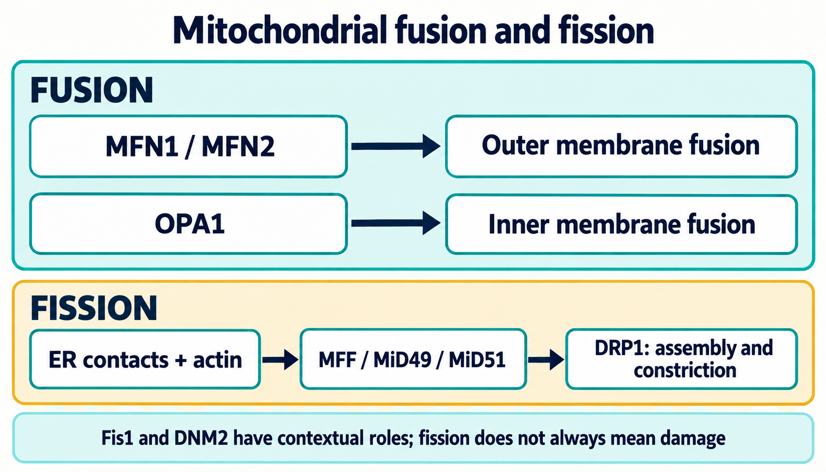
The diagram compares major fusion and fission apparatuses. Arrows describe a functional sequence rather than fixed time intervals or the sufficiency of each individual protein.
Two commonly measured DRP1 positions are called Ser616 and Ser637 in a widely used human sequence. Numbering differs between isoforms and species, so the studied protein must be checked when comparing papers. These modifications regulate assembly and localization in particular systems, but the ratio of two antibody signals is not a universal mitochondrial-health index.
On entry into mitosis, CDK1–cyclin B phosphorylates DRP1 and promotes network remodeling before cell division. This response is physiological. ERK1/2, PKCδ and CaMKII also associate with pro-fission signaling in particular stress models; phosphorylated Ser616 does not identify one specific kinase. Activity, substrate accessibility and effects on other dynamics proteins vary between cells.
In the cAMP–PKA pathway, phosphorylation of the region termed Ser637 can reduce DRP1 recruitment or activity under studied conditions. Anchoring proteins, including mitochondrial AKAPs, can spatially organize PKA. This does not amount to permanent cytosolic retention of all DRP1 or guarantee increased fusion: MFN/OPA1 machinery, lipids, energetic state and other modifications remain necessary. Some models give different results for this site; conclusions require measured fission rather than phosphorylation alone.
Calcium pulses connect metabolism, ER–mitochondria contacts and dynamics. Ca²⁺–calmodulin activates calcineurin; Cereghetti and colleagues found that it promoted DRP1 dephosphorylation and mitochondrial translocation. This is one route altering fission probability under stress, not evidence that every cytosolic calcium increase triggers irreversible fragmentation.
Signal duration and amplitude, matrix loading, calcium clearance and initial energetic reserve matter. Overload can involve permeability-transition pore opening, respiratory failure and death, but these are not automatic consequences of removing a phosphate from Ser637. Cytochrome c release additionally requires outer membrane permeability changes. Calcineurin–DRP1, mPTP and BAX/BAK are interacting yet distinct mechanisms.
Nitric oxide participates in thiol regulation and nitrosative injury; its effects depend on concentration, location and redox environment. Cho proposed that DRP1 Cys644 S-nitrosylation promotes activation, fission and neuronal injury during β-amyloid exposure. This was a testable hypothesis rather than a universal mechanism of neurodegeneration.
Bossy detected S-nitrosylated DRP1 without confirming increased enzymatic activity or Alzheimer specificity. Protein preparations, reaction conditions, SNO detection and cellular models require careful comparison. A greater-than-threefold activity increase, general protection of C644A transgenic animals and a direct causal pathway in patients cannot therefore be accepted without corresponding evidence.
These distinctions retain the topic’s scientific meaning: a redox modification may be detected, its functional effect can be condition-dependent, and clinical specificity is a separate question. Plasma nitrotyrosine measurement does not identify nitrosylation of a particular neuronal DRP1 residue.
Ubiquitin marks alter outer membrane protein availability and organize quality-control signals. Following PINK1–Parkin activation, mitofusins become ubiquitination substrates; proteasomal removal can limit re-fusion of an injured region. Gegg demonstrated PINK1/Parkin-dependent MFN1/MFN2 ubiquitination during mitophagy induction. Parkin chains are not restricted to Lys48, however, and signaling-coat formation differs from proteasomal degradation.
The membrane RING ligase MARCH5, also MARCHF5/MITOL, regulates protein-complex composition and quality, including dynamics components. Altered activity can affect mitofusins, adapters and stress responses. There is no fixed direction “more MARCH5, less DRP1, better network”: outcome depends on substrate, chain type, degradation and cell state.
The HECT ligase HUWE1, also Mule/ARF-BP1, associates with MFN2 ubiquitination in some genotoxic-stress models. This links dynamics to apoptosis experimentally, but does not make MFN2 destruction the sole requirement for BAK activation. Protein resynthesis and network remodeling can follow stress resolution; irreversibility applies to an already degraded molecule, not necessarily to the entire organelle or cell.
DRP1 SUMOylation adds another regulatory layer. Mitochondrial MUL1/MAPL and SENP proteases, including SENP5, alter SUMO attachment and complex properties. SUMO1 and SUMO2/3 need not have identical effects; membrane retention, stress contacts and subsequent fission vary by system. They cannot be reduced to a rule that every SUMO mark activates DRP1 and its removal always inhibits division.
Protein acetylation and sirtuin deacetylation connect metabolism to network regulation. Nuclear/cytosolic SIRT1 can influence PGC-1α and biogenesis programs; matrix SIRT3 acts on accessible mitochondrial substrates. These are distinct compartments. Samant reported SIRT3-dependent OPA1 deacetylation and activation in cellular and biochemical cardiac-stress experiments. Substrate accessibility and experimental context require independent consideration; such results do not follow merely from matrix SIRT3 localization. Whole-cell NAD⁺/NADH changes likewise do not establish free NAD⁺ concentration near each sirtuin.
Phosphorylation, SUMOylation and acetylation of a protein are posttranslational regulation. They should not automatically be termed epigenetic changes, which require a connection to sustained chromatin or expression regulation. The compartment logic is developed in the review of NAD⁺ consumption by CD38, PARP1 and SARM1; increasing the total cofactor pool alone does not demonstrate recovery of fusion machinery.
Fission creates regions that may differ in potential, protein composition and subsequent fate. Twig observed connections between division, selective re-fusion and autophagic removal in studied cells. A lower-potential daughter could return to the network less readily and undergo degradation more often. This is an important damage-segregation model, not evidence that most divisions universally yield one healthy and one defective organelle.
Fission also supports mitochondrial proliferation and distribution, while transient depolarization need not mean irreversible injury. Demonstrating asymmetry requires observation before and after division, appropriate potential-sensitive dyes and controls for size, dye loading and phototoxicity. One fluorescence image cannot identify mitochondrial-DNA mutation accumulation, and a fixed potential threshold is not a universal selection criterion.
Fusion can provide functional complementation, but does not chemically remove an oxidized protein or automatically repair a mutant genome. Damage removal requires proteolysis, component renewal or completed autophagic flux. Excess fusion suppression is also harmful: cells must maintain content exchange and the function of the remaining pool.
With preserved energetics, PINK1 interacts with TOM/TIM import machinery, undergoes processing involving mitochondrial proteases including PARL, and is rapidly removed. This differs from a model in which the entire protein continuously enters the matrix and is simply cleaved there. Import, transmembrane-segment positioning, proteolytic stages and product export determine PINK1’s short lifetime on functional mitochondria.
When potential-dependent import is substantially impaired, full-length PINK1 stabilizes in a complex at the outer membrane, allowing its catalytic region to interact with cytosolic substrates. Narendra demonstrated selective stabilization on impaired mitochondria and its connection to Parkin activation. Depolarization duration and agent matter: uncoupler treatment in culture does not automatically reproduce every human mitochondrial stress.
PINK1 phosphorylates ubiquitin at Ser65 and the corresponding serine in Parkin’s UBL domain. These are distinct substrates. Kondapalli’s 2012 paper examined phosphorylation-mediated Parkin activation; primary evidence for ubiquitin phosphorylation and its Parkin-activating role includes Koyano’s 2014 study. Confusing the titles turns separate cascade steps into an inaccurate source attribution.
Phosphoubiquitin binding and phosphorylation help release autoinhibition of the RBR E3 ligase Parkin. Parkin transfers ubiquitin to outer membrane proteins; newly created ubiquitin substrates can be phosphorylated by PINK1, amplifying the local signal. The result is positive feedback rather than a single mark on a mitofusin.
MFN1/2, transport components and other membrane proteins can become substrates. Ubiquitination affects fusion, motility and recognition by autophagy receptors. Proteasomal removal of some outer membrane proteins differs from subsequent lysosomal organelle removal. MFN degradation and reduced transport may support local isolation, but do not demonstrate that the whole mitochondrion has already been destroyed.
The ubiquitin coat connects mitochondria to selective-autophagy receptors. OPTN and NDP52 are important in studied PINK1/Parkin systems; TBK1 regulates their functional interactions and response amplification. Receptors recruit phagophore machinery and interact with LC3/GABARAP-family proteins. The isolation membrane grows around the cargo, closes into an autophagosome and subsequently interacts with the lysosomal compartment.
p62/SQSTM1 can participate in aggregation of ubiquitinated material and particular autophagic responses, but is not the only obligatory bridge from all mitochondria to LC3. Lazarou demonstrated distinct receptor functions, precluding one universal “p62–LC3” arrow.
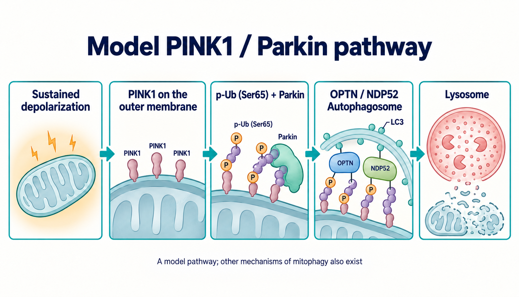
The diagram depicts a model route from sustained depolarization to lysosomal degradation. Other mitophagy mechanisms, including receptor pathways without obligatory Parkin, operate in different tissues and conditions. Even absence of Parkin does not mean absence of all mitochondrial renewal.
Mitophagy confirmation requires flux measurements: mitochondrial-material delivery to an acidic compartment and degradation over time, with lysosomal-function controls. LC3 puncta, membrane-associated Parkin or reduced mitochondrial mass alone are ambiguous. Lysosomal blockade can increase marked organelles while reducing completed removal; mass decline may reflect injury, cell death or reduced biogenesis.
Pathogenic MFN2 variants are an established cause of some axonal Charcot–Marie–Tooth type 2A cases. Züchner linked a dynamics protein to inherited neuropathy. Inheritance is often autosomal dominant, but other genetic situations occur; severity, onset and additional features differ among variants and families.
A long axon relies on mitochondrial delivery to high-demand regions and organelle return or local renewal. Miro, TRAK, kinesins and dynein organize microtubule transport; MFN2 relates to this organization in addition to fusion and ER contacts. Impaired movement, distribution or content exchange can reduce distal energetic reserve and calcium buffering.
Individual MFN2 variants nevertheless produce different functional phenotypes. Not every variant completely disrupts KIF5 association, removes mitochondria from all distal axons or inhibits GTPase activity identically. Mechanistic assessment compares genetics, cellular models, time-resolved transport, fusion and bioenergetics. Improving one parameter in patient-derived culture is not equivalent to restoring human nerve function.
Pathogenic OPA1 variants cause dominant optic atrophy; Alexander established the primary genetic association. Retinal ganglion cells and their axons are particularly vulnerable to disrupted energetics, crista organization, mitochondrial-DNA maintenance and transport. Human genetics here supports protein causality more strongly than a shape change alone in an acquired disorder.
Many variants reduce functional OPA1, while others change protein properties; severity and presentation age vary. DOA-plus phenotypes may include additional neurological or systemic abnormalities, including hearing, motor or muscular features. They are not identical in all carriers.
Impaired inner fusion and crista architecture can worsen respiration and vulnerability to death, but no single compulsory sequence of “crista opening, immediate cytochrome c release, apoptosis” describes every patient. Clinical evaluation, variant-specific interpretation and alternative causes of visual dysfunction remain necessary.
Altered dynamics, transport and mitochondrial energetics occur in neurons and Alzheimer models. Manczak examined β-amyloid–DRP1 interactions and impaired dynamics; tau and signaling-kinase observations add to the picture. Organelle shortening can accompany impaired synaptic energy supply and calcium regulation.
Postmortem tissue, culture and amyloid-expression models nevertheless answer different questions. A DRP1–amyloid correlation does not prove hyperfission is invariably the earliest sufficient disease event. Causality and temporal ordering require perturbation, longitudinal observation and functional outcomes. The SNO-DRP1 dispute also prevents presenting one redox mechanism as an established explanation in patients.
Pathogenic PINK1 and PRKN variants associate with inherited parkinsonism, providing a strong basis for studying mitochondrial quality control. PINK1–Parkin disruption alters responses to particular injuries, but does not switch off all mitophagy. Basal turnover and receptor-dependent responses can persist; compensation depends on tissue and conditions.
Particular α-synuclein forms interact with the import receptor TOM20 and impair protein import. Di Maio combined biochemical and cellular experiments with postmortem-tissue observations. This does not mean every α-synuclein monomer blocks TOM20 identically or that the pathway explains every disease case.
Mitochondrial injury can contribute to inflammatory signaling, including mitochondrial-DNA release and cGAS–STING or NLRP3 pathways. Each requires its own activation conditions and direct measurements. They cannot form a compulsory linear cascade from every Parkin abnormality to persistent neuroinflammation and dopaminergic-neuron death.
In metabolic models, network fragmentation can accompany lipid overload, respiratory changes and altered insulin responses. Shape does not replace measurement of insulin-receptor-to-IRS–PI3K–AKT signaling, glucose transport or tissue insulin sensitivity. A single IRS1 serine phosphorylation, including frequently discussed rodent Ser307, is species- and context-dependent rather than universal evidence of insulin-signal inhibition.
In β-cells, metabolism links glucose entry to ATP/ADP, KATP-channel closure, plasma-membrane depolarization, calcium influx and insulin secretion. Dynamics abnormalities can affect this response, but organelle length alone does not determine secretion. Energetics, calcium dynamics and insulin release require measurement.
Ischemia–reperfusion involves energetic depletion, calcium overload, redox changes, ROS formation on oxygen return and membrane permeability transition. DRP1 and network remodeling contribute in some injury models, but are not the sole infarct-size regulators. mdivi-1 protection needs particular caution: complex I effects can modify ROS independently of direct DRP1 inhibition.
mdivi-1 is a quinazolinone derivative identified in the fission-machinery study by Cassidy-Stone. It has been widely used experimentally as a fission inhibitor, but Bordt demonstrated reversible effects on mitochondrial complex I. Reduced ROS or cell protection therefore does not establish direct selective inhibition of human DRP1. Electron-transfer inhibition is also not identical to mild uncoupling: respiration, potential and proton conductance distinguish them.
Causal analysis benefits from independent genetic DRP1 perturbation, respiratory-effect controls and restoration of protein function. If an effect persists without the proposed target, it needs another mechanistic explanation. Improved network shape or reduced animal-model injury does not establish a human treatment regimen.
P110 is an experimental peptide studied by Qi to limit DRP1–Fis1 interaction in stress models. The construct includes DLLPRGT and is used in some experiments with a cell-penetrating TAT fragment. Studies describe reduced pathological fragmentation and injury in particular systems. Claimed selectivity must be evaluated against method, concentration and exposure duration; complete preservation of every physiological division and mitophagy event cannot be guaranteed in all tissues. These data do not constitute a clinical P110 prescribing protocol.
M1 was studied as a mitochondrial fusion promoter in a phenotypic cellular screen. Wang demonstrated the possibility of chemically changing the network. Chemical identification of the hydrazone derivative is useful for reagent identity, but does not establish one direct protein target. Restoring tubular morphology during partial machinery impairment does not mean correcting every MFN1, MFN2 or OPA1 defect.
Franco proposed mitofusin activation through conformational-interaction changes; Rocha studied MFN2 agonists in preclinical CMT2A models. Compounds labeled B-S8 or 8015 in particular programs must be distinguished by structure and experiment rather than grouped as one drug. Open conformation, trans interaction, transport and functional fusion are connected but separately testable properties.
Improved organelle transport or animal behavior is of translational interest. Clinical conclusions nevertheless need nerve exposure, duration, toxicity, MFN2-variant and sustained functional-outcome data. A selected mouse model does not establish reversal of paralysis in patients or identical responses of all pathogenic variants. Continuous fusion stimulation is not invariably beneficial either: it may hinder separation of regions requiring removal.
Activity changes energy demand and mitochondrial-renewal programs. AMPK, p38 MAPK and PGC-1α participate in adaptation, but an acute response differs from weeks of training. Experimental exercise can activate fission and mitophagy alongside subsequent functional improvement. Increased DRP1 after exercise is therefore not automatically damage, and elongation alone does not demonstrate beneficial training.
Moderate aerobic activity, often called zone 2, has physiological meaning, but different zone definitions do not exactly match one lactate threshold. It cannot be prescribed as the sole proven intensity for restoring everyone’s MFN2/OPA1. Total volume, tolerance, baseline fitness and measured functional changes, including aerobic performance, matter; expression of one gene does not replace outcomes.
Daily organization of sleep, eating and activity affects the metabolic background. General links between circadian regulation and energetics nevertheless do not establish an hourly schedule of human DRP1 activation or obligatory hyperfusion. Such claims require tissue- and time-specific measurements.
Gomes described nutrient-deprivation elongation through cAMP–PKA and DRP1 regulation experimentally. This does not establish a universal 14–16-hour fasting boundary after which a human network necessarily becomes protected. Medium composition, tissue, duration and lysosomal-removal capacity modify the response. Calorie restriction, 16/8 and OMAD are also different interventions; a mechanistic hypothesis does not establish personal benefit or tolerability.
A substance may change redox state, autophagy markers or transcription without directly activating MFN2 or inhibiting DRP1. The table retains the supplied manuscript’s main groups, but does not label proposed doses as a clinical therapeutic window: no such range is established for treating a dynamics disorder.
| Substance | Studied mitochondrial association | What the mechanism does not establish |
|---|---|---|
| Urolithin A | Ellagitannin metabolite; model mitophagy and molecular human-study measures | Marker changes do not prove obligatory PINK1/Parkin, fusion or strength recovery in everyone |
| Spermidine | Polyamine; experimental autophagy and acetylation regulation | Model cardioprotection and longevity do not establish clinical OPA1 restoration |
| Resveratrol / pterostilbene | Redox/signaling effects; SIRT1–PGC-1α in some models | Direct SIRT1 activation and site-specific DRP1 inhibition depend on the system; one substance’s effect does not transfer to another |
| Coenzyme Q10 / ubiquinol | Electron transfer from complexes I/II to III; membrane redox balance | No universal direct OMA1/YME1L block or restoration of dynamics under every deficiency is established |
| Astaxanthin | Lipid-membrane interaction and antioxidant effects in models | Bilayer orientation does not prove preservation of all cristae, OPA1 complexes or mPTP closure in humans |
Andreux studied urolithin A in humans and found molecular changes compatible with mitochondrial effects. These differ from direct observation of completed mitophagy in every tissue or demonstrated treatment of neuropathy or age-related weakness. Trial results depend on population, duration and endpoint; a marker increase is not a strength gain.
Eisenberg’s spermidine study supports experimental interest in autophagy and cardioprotection. Reducing its effects to direct OPA1 deacetylation through EP300 would overstate the evidence: enzyme targets, general autophagic flux and crista architecture require different measurements. Similarly, resveratrol’s SIRT1 effects depend on substrate and assay conditions, while PGC-1α or ERK changes do not automatically transfer to clinical network dynamics.
Coenzyme Q10 participates in the respiratory chain, but “protection from oxidative degradation of zinc ions” in OMA1/YME1L is chemically inaccurate and does not justify a targeted action. For astaxanthin, membrane models must be separated from organelle function and clinical outcomes, as discussed in the article on astaxanthin, lipid bilayers and peroxidation. Together these mechanisms do not establish a combined fusion-restoration protocol.
Inherited dynamics disorders are assessed through clinical phenotype, family history and genetics. Panels or broader sequencing may include MFN2, OPA1, DNM1L, MFF, PINK1 and PRKN when appropriate to the diagnostic question. A variant requires classification, phenotype matching and sometimes segregation analysis; a variant of uncertain significance does not itself confirm disease.
Research models use live fusion/fission imaging, L-/S-OPA1 analysis, potential dyes and oxygen-consumption measurements. OCR measures respiration under defined conditions, while ECAR measures extracellular acidification influenced by more than glycolysis. These methods test mechanisms but do not constitute a universal clinical panel from one blood mononuclear-cell sample. An accessible cell need not represent neurons, retina or muscle.
3-Nitrotyrosine, malondialdehyde and circulating cell-free mitochondrial DNA are nonspecific. Values depend on material source, sample handling, inflammation and cell injury. They do not establish a mitophagic collapse or identify dysfunction of a particular DRP1 residue, OMA1 protease or mitofusin.
An intervention should specify its intended effect: completed division, matrix-content exchange, transport, autophagic flux, respiratory reserve or tissue function. Several concordant methods and independent perturbation of the proposed target support stronger conclusions than one elongated-network image. Clinical research additionally needs patient-relevant outcomes and safety assessment.
MFN2, OPA1 and DRP1 show how organelle shape connects to energetics, transport and renewal. Maximizing fusion or permanently suppressing fission cannot be the objective. P110, mitofusin agonists and metabolic interventions each need separate evaluation; combining preclinical mechanisms does not create an already established treatment protocol. A functional cell retains the ability to remodel its network and remove damage according to tissue needs.
This is a critical narrative review based on the supplied manuscript and its primary literature. During local editorial preparation on 8 October 2026, primary records were checked specifically for disputed mechanisms and incorrect bibliographic associations: MFN2–ER contacts, DNM2 necessity, SNO-DRP1, mdivi-1 complex I effects, Mgm1 versus OPA1, PINK1 substrates, autophagy receptors, OPA1 acetylation and selected nutrient/exercise models. No new exhaustive verification of every supplied bibliography link is claimed.
Structural constructs, cellular and animal models, human genetics and biomarker trials are distinguished. Universal numerical thresholds, unconfirmed therapeutic doses and definitive clinical promises were not transferred from mechanistic diagrams to conclusions. This is not a systematic search, GRADE assessment or external peer review. Diagrams are explanatory models and do not replace experimental flux or tissue-function measurement.
No. Fission contributes to mitosis, transport and adaptation; function and subsequent fate, rather than shape alone, determine its condition.
No. Complex I effects have been demonstrated, so protection requires an independent mechanistic check.
No. Other receptor and tissue pathways exist; Parkin recruitment also does not confirm completed lysosomal degradation.
Combining experimental mechanisms does not demonstrate clinical effectiveness. Outcomes and safety require appropriate studies.