
Mitochondrial calcium uniporter MCU: from ER microdomains to energy metabolism and overload
How MCU, MICU and EMRE admit Ca²⁺ from ER microdomains, support dehydrogenases and interact with NCLX. Structures, equations, overload and limits of pharmacological and nutritional inference.
Calcium connects cellular work to the mitochondrial energy response. During muscle contraction, neuronal signalling or secretion, brief Ca²⁺ rises can accelerate production of reducing equivalents in the matrix. Calcium must cross both mitochondrial membranes, and uptake must be coordinated with efflux, buffering and ATP demand.
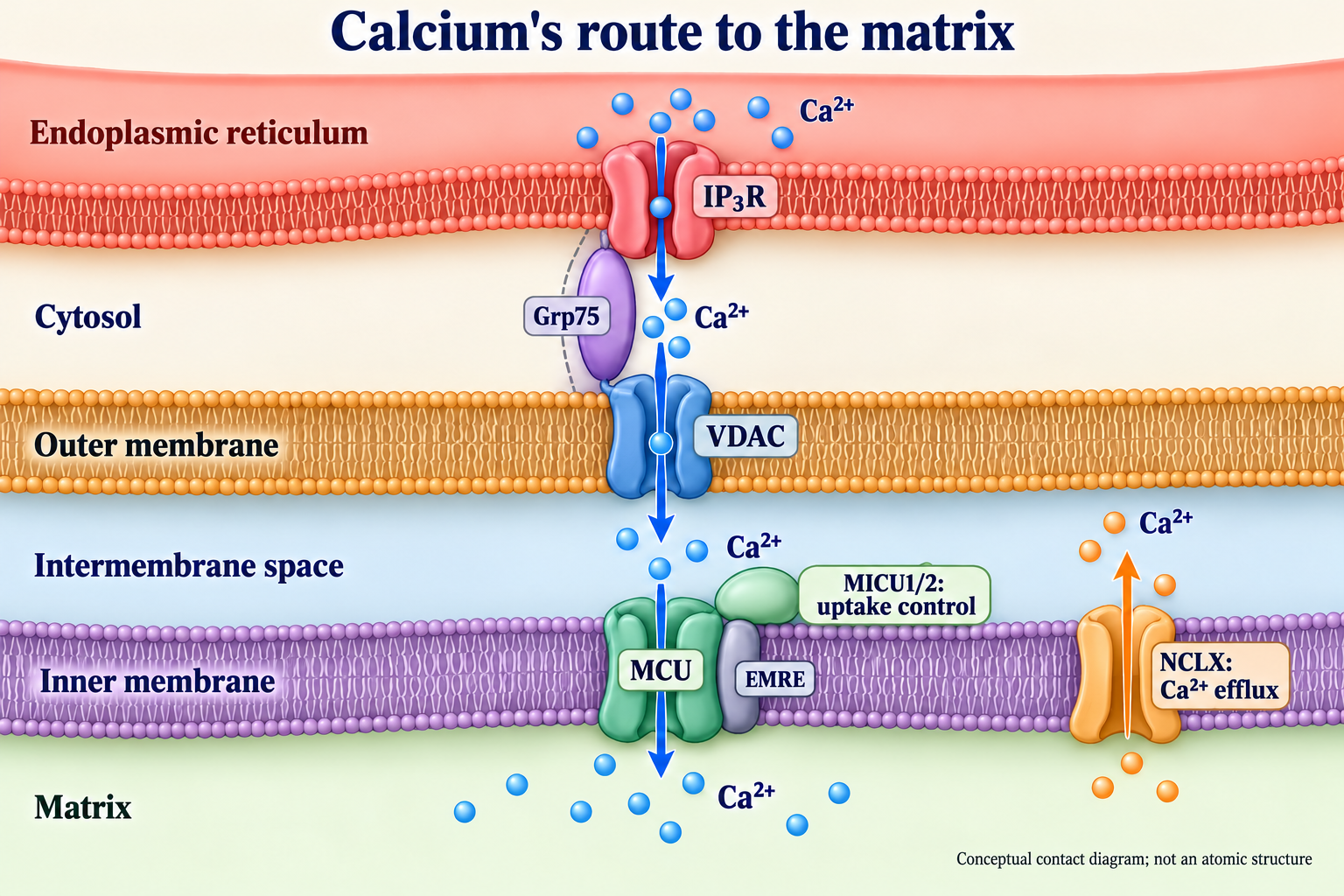
The mitochondrial calcium uniporter, MCU, forms a channel in the inner membrane. MICU proteins and EMRE, contacts with the endoplasmic reticulum, NCLX and the respiratory chain determine the result together. Sustained overload under adverse conditions increases the risk of damage; a useful signal cannot be described by calcium abundance or MCU expression alone.
Electrochemical foundations
Calcium as a signal and a load
Resting cytosolic free Ca²⁺ is often around 50–100 nM, whereas extracellular ionised calcium is in the millimolar range. These reference values vary with the cell and measurement conditions. Channels, pumps and buffers shape pulses, so the average concentration does not describe a mitochondrion's local exposure.
Free and total matrix calcium must be distinguished. Binding to proteins, metabolites and phosphate allows accumulation without a proportional rise in the free fraction. This does not mean that any concentration above the submicromolar range immediately causes irreversible salt deposition or cell death.
Inner-membrane potential
The respiratory chain creates a proton electrochemical gradient. The matrix is usually negative relative to the intermembrane space; values around −160 to −180 mV are often quoted, but depend on tissue, workload and method. MCU uses the electrical component to admit positively charged Ca²⁺ and does not itself hydrolyse ATP.
With Δψ = ψmatrix − ψintermembrane space and ΔpH = pHmatrix − pHintermembrane space, the electrochemical-potential change for proton entry is:
F is the Faraday constant, R the gas constant and T absolute temperature. A 0.18-V difference across an illustrative 5-nm membrane corresponds to about 3.6 × 10⁷ V/m, or 360 kV/cm. This estimates a field across a nanoscale distance; it establishes neither a whole-body voltage nor a particular transport rate.
Interpreting the Nernst calculation
For a divalent cation at ideal equilibrium:
At 37 °C, the denominator in the base-10 form for a monovalent ion is approximately 61.5 mV. For Ca²⁺ at Δψ = −180 mV, the activity ratio is about 7 × 10⁵. Applying an external free concentration of 100 nM would give a hypothetical internal value near 70 mM.
This illustrates the strong driving force, rather than predicting normal free matrix Ca²⁺. A living mitochondrion is outside this equilibrium: channel regulation, efflux, buffering and changing voltage all matter. Replacing those processes with one equilibrium number creates a misleading picture of permanent overload.
From physiology to molecular identification
Calcium uptake by isolated mitochondria was studied in the 1960s. Kirichok and colleagues' 2004 mitoplast recordings demonstrated a highly selective calcium channel. MICU1 was identified in 2010; independent studies by Baughman and De Stefani identified pore-forming MCU in 2011. Electrophysiology, genetics and structural methods answer complementary questions and cannot substitute for one another.
Channel architecture and its regulators
The MCU tetramer and membrane topology
The human MCU precursor contains 351 amino acids. After import, the protein forms an inner-membrane complex with a matrix-facing N-terminal domain, two transmembrane segments and helical assembly elements. Four MCU subunits form the pore; TM2 faces the conducting pathway, while TM1 lies closer to surrounding lipids. The intervening loop faces the intermembrane space.
Structure 6DNF is fungal MCU from Cyphellophora europaea, rather than the human holocomplex; Baradaran 2018 reported 3.2-Å resolution. Human MCU–EMRE in Wang 2019 structure 6O58 is a dimer of channels at 3.8 Å and lacks the complete MICU complement. Fan 2020 structure 6WDN is the human MCU–EMRE–MICU1–MICU2 holocomplex at low Ca²⁺, resolved at 3.2 Å. Species, composition and state are essential distinctions.
The DIME filter and selectivity
The conserved DIME sequence comprises Asp261, Ile262, Met263 and Glu264 in the numbering of the human precursor. Acidic residues form important elements of the selectivity filter. Their arrangement helps explain Ca²⁺ preference and ruthenium blockade, but an electrostatic sketch alone does not fully explain conductance.
The ion interacts with both the filter and its hydration shell. Multi-ion models address the combination of tight binding and rapid conduction. Hydration energies depend on the thermodynamic convention used; they cannot directly establish universal MCU binding constants or prove a single knock-on mechanism.
MCUb and complex composition
MCUb, encoded by MCUB/CCDC109B, is an MCU paralogue that can reduce calcium conductance when incorporated into the complex. This is experimentally demonstrated regulation of channel composition, rather than a compulsory two-MCU/two-MCUb arrangement in every tissue. MCUb abundance and complex remodelling vary with development, injury and adaptation.
Pore-region sequence differences are important, but their effects require mutagenesis and current measurements. A transcript ratio cannot by itself determine cardiac flux, and increasing MCUb does not establish protection in a patient.
EMRE and coupling to MICU
EMRE, the SMDT1 product, is a small single-pass membrane protein required for effective human MCU transport. Its interactions support a functional complex; the acidic C-terminal region on the intermembrane side contributes to MICU coupling. Sancak 2013 showed why identifying the pore alone did not complete the mammalian mechanism.
EMRE dependence is not universal across organisms. Some fungal MCUs function without this partner. Structural comparison must account for evolutionary architecture rather than assume an identical protein complement in all mitochondria.
Lipids and cardiolipin
The inner membrane provides a lipid environment as well as the electrical gradient. Cardiolipin influences the organisation of several mitochondrial proteins and membranes; lipid changes can affect assembly and transport. Its broader effects do not prove that every disturbance of cardiolipin metabolism acts exclusively through MCU.
Discussion of Barth syndrome, lipid oxidation or membrane instability must also consider respiratory complexes, cristae and other carriers. Calling cardiolipin “MCU glue” is at most a rough metaphor, rather than a measurement of the specific complex.
| Component | Location | Main function | Important limitation |
|---|---|---|---|
| MCU | Inner membrane; NTD in matrix | Ca²⁺ uptake pore | Flux depends on voltage and regulators |
| MCUb / MCUB | Inner membrane | Composition control and reduced conductance | No universal ratio to MCU |
| EMRE / SMDT1 | Inner membrane | Functional support and MICU coupling | Requirement varies by species |
| MICU1 | Intermembrane side | EF-hand sensor and uptake regulation | Threshold depends on complex and conditions |
| MICU2 | Intermembrane side | Regulation together with MICU1 | Not an independent pore |
| MICU3 | Intermembrane side | Enhanced response in some tissues | Not a universal MICU2 replacement |
| NCLX / SLC8B1 | Inner membrane | Na⁺/Ca²⁺ exchange and Ca²⁺ efflux | Depends on ionic gradients |
| VDAC1 | Outer membrane | Access for Ca²⁺ and metabolites | Not an inner-membrane channel |
| IP₃R | ER membrane | ER calcium release | Transfer depends on contact geometry |
| Grp75 / HSPA9 | Several cellular pools | Chaperone; contact-complex participation | Not another calcium pore |
| Cyclophilin D / PPIF | Matrix | Regulation of mPTP sensitivity | Molecular composition of mPTP remains under investigation |
MICU proteins and calcium sensing
EF-hand sensors at low exposure
MICU1 and MICU2 contain calcium-binding EF-hand domains. At low Ca²⁺ they restrict unwanted uptake; a rise in local concentration changes their state and facilitates transport. This coordinates uptake with a signal without implying absolutely zero basal flux below one universal threshold.
Complex composition, Mg²⁺, voltage, tissue and measurement method differ between experiments. Statements such as “closed below 1 µM” or “opens precisely at 10 µM” describe particular systems and are not clinical boundaries.
The heterodimer and channel contact
MICU1 and MICU2 form a regulatory pair. Intersubunit interactions, including a disulfide bond, organise this pair; residue numbering varies by species and precursor versus mature sequence. Low- and high-calcium structures show rearrangement of MICU relative to the pore.
Positively charged MICU surfaces interact with acidic entry regions and complex partners. A list of Lys/Arg residues from one model cannot be transferred indiscriminately to every structure. Nor is regulation solely a mechanical lid: accessibility, sensitivity and conductance are related but different properties.
Cooperativity and the Hill equation
A sigmoidal calcium–flux relationship can be approximated by:
C is Ca²⁺ concentration in the experimental compartment, K₀․₅ the half-response concentration and n an empirical Hill coefficient. Values near n = 2.5–3 and micromolar K₀․₅ characterise some measurements. The coefficient is not automatically the number of bound ions and does not establish a protein phase transition.
MICU3 and tissue-specific tuning
MICU3 can partner with MICU1 and enhance the mitochondrial response to smaller calcium signals. This is especially relevant to neurons with rapidly changing energy demand. Patron 2019 demonstrated such regulation in experimental systems.
MICU3 is a distinct protein rather than an alternative MICU2 isoform. Its presence does not imply an identical several-fold enhancement throughout neurons and muscle. Contacts, buffers, pulse frequency and mitochondrial condition also shape the response.
Inherited regulatory disorders
Biallelic human MICU1 defects are associated with myopathy and neurological features including developmental delay and movement abnormalities. They demonstrate the physiological importance of threshold and transport dynamics. A regulator-loss cell model helps investigate mechanisms, but does not specify an identical phenotype for every patient.
Chronic dysregulation cannot be reduced to obligatory calcification of all mitochondria. Assessment of an inherited disorder requires the variant, phenotype and functional evidence; ordinary fatigue alone does not diagnose a MICU defect.
ER–mitochondrial contacts and local Ca²⁺ delivery
Contact geometry and MAM
Membranes at ER–mitochondrial contacts are often separated by tens of nanometres. A 10–30-nm range is illustrative for some contacts and varies with cell and method. MAM originally denotes a mitochondria-associated membrane fraction; biochemical isolation and microscopic definitions of contact do not coincide completely.
Contacts participate in lipid exchange, autophagy and signal organisation. Their area is not a fixed proportion of mitochondrial surface across tissues. More contacts do not necessarily improve energy supply: prolonged or excessive transfer can increase load.
IP₃R, Grp75 and VDAC
IP₃ receptors release calcium from the ER. Grp75/HSPA9 participates in functional coupling between IP₃R and outer-membrane VDAC1. Calcium then reaches the intermembrane space and crosses the inner membrane through MCU. These are separate membranes and proteins, rather than a single IP₃R–Grp75–VDAC–MCU pipe.
Grp75 is a chaperone with several cellular pools, including the matrix pool; contact-associated protein does not make all Grp75 exclusively cytosolic. In muscle, sarcoplasmic-reticulum calcium released through ryanodine receptors also matters. One configuration does not encompass every type of contact.
Microdomains versus average cytosolic calcium
Near an open release channel, local calcium can briefly reach tens of micromolar while the cytosolic average remains much lower. This explains how a micromolar-sensitive MCU responds to a cellular signal. A 20–50-µM range illustrates some settings and is not a property of every contact.
Diffusion, calcium binding and channel-closing kinetics determine microdomain duration. Local exposure, distance and pulse shape can therefore be more informative than one averaged concentration. Microdomains obey ordinary physics and do not remove thermodynamic constraints.
MFN2, PACS2 and other organisers
MFN2 participates in mitochondrial dynamics and ER-contact regulation. Its tethering role has been debated in studies with differing results, including de Brito 2008, Filadi 2015 and Naon 2016. Cell types, methods and definitions of proximity require caution with the claim that MFN2 removal always reduces contacts.
PACS2 and BAP31–FIS1 interactions also relate to interorganelle organisation and stress. They do not form one compulsory scaffold with a fixed distance. Charcot–Marie–Tooth disease type 2A associated with MFN2 variants cannot be explained solely by failed calcium transfer; transport, fusion and other mitochondrial functions matter.
Calcium and the energy response
PDP1 and pyruvate dehydrogenase
The pyruvate dehydrogenase complex links glycolysis to the tricarboxylic-acid cycle:
Calcium regulates it primarily through phosphatase PDP1, promoting E1α dephosphorylation and complex activation. PDK1–4 kinases act in the opposite direction. Three familiar E1α sites are Ser293/300/232 in the precursor or Ser264/271/203 after targeting-peptide removal; mixing conventions produces apparent contradictions.
Mg²⁺ participates in phosphatase catalysis, while Ca²⁺ influences PDP1 association with the complex. PDP2 has different regulatory properties and is not an equivalent calcium switch. Activation constants and binding changes measured in particular experiments are not universal. E2-bound lipoyl groups must also be distinguished from oral lipoic acid, as discussed in the article on R-alpha-lipoic acid and PDC.
NAD-dependent isocitrate dehydrogenase IDH3
IDH3 produces 2-oxoglutarate:
It differs from NADP-dependent IDH2. Calcium and adenine-nucleotide regulation help coordinate the cycle with workload; ADP interaction and altered apparent substrate affinity are important. The subunit organisation of human IDH3 must be described with reference to the structural unit and assembly state rather than a universal formula for every species.
Published shifts of apparent Kₘ from millimolar to submillimolar values refer to particular conditions. They do not mean that increasing calcium always multiplies flux by a fixed factor: substrates, NADH/NAD⁺, ATP/ADP and downstream reactions also matter.
The 2-oxoglutarate dehydrogenase complex
OGDH/OGDC performs another oxidative decarboxylation:
Calcium can enhance substrate responsiveness; classical studies described lower apparent Kₘ. Unlike PDC, this is not the PDK/PDP switch. Absence of that particular scheme does not imply absence of all post-translational modifications or other regulation.
A standard ΔG°′ near −33.5 kJ/mol is a thermodynamic reference, whereas actual ΔG depends on metabolite activities. NADH and succinyl-CoA can constrain flux, and the complex is redox-sensitive. It is therefore not an unconditional metabolic engine controlled only by calcium.
ARALAR, CITRIN and the malate–aspartate shuttle
ARALAR/SLC25A12 and CITRIN/SLC25A13 are inner-membrane aspartate/glutamate carriers, not adenine-nucleotide carriers. Their regulatory EF-hand regions face the intermembrane side and can respond to calcium outside the matrix. Calcium control of metabolism is thus not limited to ions that have passed through MCU.
The malate–aspartate shuttle transfers reducing equivalents from cytosolic NADH through metabolite exchange. NADH itself does not cross the inner membrane as ARALAR/CITRIN cargo. Tissue distribution and availability of shuttle partners determine its contribution to respiration.
Respiration, ATP synthase and feedforward regulation
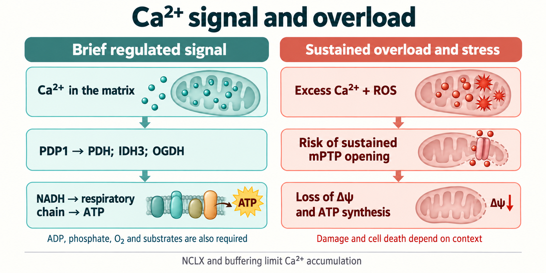
Increased NADH production can support electron flow and ATP synthesis when oxygen, ADP, phosphate and substrates are available. This feedforward response complements feedback from energy demand. Calcium also activates ATP-consuming processes; a useful response matches production to consumption.
This does not establish a universal direct mechanism in which Ca²⁺ displaces IF1 and invariably accelerates the ATP-synthase rotor. IF1, pH, potential and enzyme direction have their own regulation. Maintenance of ATP during a several-fold workload increase is an experimental observation, not obligatory behaviour in every cell.
| Target | Calcium-sensing location | Energy relationship | Unsupported automatic inference |
|---|---|---|---|
| PDP1 → PDC | Matrix | E1α dephosphorylation and pyruvate oxidation | All PDP forms are equally sensitive |
| IDH3 | Matrix | Regulation of an NADH-producing reaction | IDH2 uses the same mechanism |
| OGDH / OGDC | Matrix | Altered response to 2-oxoglutarate | PDK/PDP regulates it as it does PDC |
| ARALAR / CITRIN | EF-hands on intermembrane side | Malate–aspartate shuttle | They carry NADH or adenine nucleotides |
| Respiration and ATP synthase | Integrated system response | Matching reducing equivalents to demand | Calcium replaces ADP, O₂ or substrates |
Overload, buffering and injury
Ischaemia, reperfusion and excitotoxicity
During ischaemia, oxygen limitation reduces ATP production and disrupts ion homeostasis. Changes in Na⁺, pH and membrane potential can raise cytosolic calcium load; in some settings the plasma-membrane Na⁺/Ca²⁺ exchanger reverses. This is a context-dependent mechanism, rather than one fixed calcium concentration in all patients.
Restoring blood flow changes pH, respiration, Δψ and ROS production together. Recovery of the driving potential can favour uptake while cellular load persists. Excessive NMDA-receptor activation and other sources of Ca²⁺ contribute to neuronal excitotoxicity. Duration, localisation, buffering and energetics determine overload.
Phosphate buffering and chemical-model limits
Phosphate helps retain calcium in the matrix. Possible calcium-phosphate stoichiometries include:
These equations balance mass and charge, but do not establish that mitochondrial granules always constitute mature hydroxyapatite. Amorphous complexes influenced by pH, Mg²⁺ and organic constituents are often considered. Buffering can lower the free fraction while increasing total content. “99% bound calcium” is not a universal statement without measurement conditions.
mPTP and Cyclophilin D
Sustained opening of the mitochondrial permeability-transition pore, mPTP, can disrupt inner-membrane selectivity, reduce Δψ and impair ATP synthesis. Calcium, oxidation and other factors change transition probability. Cyclophilin D, encoded by PPIF, regulates sensitivity; it is not established as a compulsory pore-forming subunit whose sole role is direct calcium sensing.
ANT- and ATP-synthase-associated models, including different synthase assemblies, remain under investigation. Transient permeability events are also distinct from sustained pathological opening. An operational description involving molecules near 1.5 kDa does not establish one precise pore geometry.
From membrane injury to apoptosis
Persistent inner-membrane disruption can cause swelling and outer-membrane injury. Cytochrome c release, association with Apaf-1 in the presence of nucleotides and apoptosome formation activate caspase-9 and executioner caspases-3/7. Cristae organisation and cytochrome c–cardiolipin interactions influence protein availability.
Smac/DIABLO and Omi/HtrA2 interact with apoptosis-inhibitor systems; AIF and endonuclease G participate in some caspase-independent pathways. However, BAX/BAK-mediated outer-membrane permeabilisation can occur without obligatory mPTP opening. Not all apoptosis follows a single MCU → mPTP → cytochrome c sequence.
Energy failure, necrosis and inflammation
After potential loss, ATP synthase can reverse and consume ATP under certain conditions; IF1 and pH affect this behaviour. Depletion does not always occur within a fixed number of minutes. Severe energy deficit often favours necrotic outcomes, but does not absolutely determine death mode.
Injured cells can release mitochondrial DNA, HMGB1 and other danger signals, engaging inflammation. Baines and Nakagawa's 2005 Cyclophilin-D-loss studies distinguished some necrotic and apoptotic mechanisms. They argue against a universal model in which blocking one regulator prevents every form of cell death.
NCLX: efflux and signal duration
An exchanger rather than an ATP-driven pump
NCLX/SLC8B1 mediates inner-membrane Na⁺/Ca²⁺ exchange and can use Li⁺ experimentally. A commonly used electrogenic model has three Na⁺ entering for one Ca²⁺ leaving:
Here in means matrix and out the intermembrane side. This direction transfers one net positive charge inward. Flux depends on sodium, calcium and voltage; other transport processes, including proton exchange, help maintain the sodium gradient. NCLX does not directly use ATP as a pump.
Flux balance and buffering
MCU is a channel, whereas NCLX cycles between carrier conformations, so their kinetics differ. A universal 20–50-fold speed ratio or fixed turnover for every mitochondrion is not established. Total calcium balance can be written:
With consistent flux and volume normalisation, the relation to free calcium involves β = dCₜ/dCfree. Stronger buffering means a smaller free-concentration change for the same total-content change. Incomplete recovery between pulses can produce accumulation without an unusually large individual signal.
Heart and nervous system
Luongo 2017 found that conditional NCLX deletion in the adult mouse heart caused severe dysfunction and early death; increased expression in other experiments limited overload. This demonstrates the importance of efflux in a specific model rather than establishing a human heart-failure treatment.
NCLX alterations and PINK1-related signalling are investigated in neurodegeneration. Experimental links do not make NCLX deficiency the single established cause of Alzheimer's or Parkinson's disease. Translation requires delivery, tissue selectivity, safety and controlled clinical outcomes.
Pharmacological tools and nutritional hypotheses
Ruthenium red, Ru360 and Ru265
Ruthenium red has long been used to limit mitochondrial Ca²⁺ uptake, but has additional targets. Ru360 is more selective in several systems; low plasma-membrane permeability complicates intact-cell studies. Its permeability cannot be called absolutely zero under every preparation and condition.
Ru265 was developed as a more cell-permeable ruthenium tool. Woods 2019 investigated its properties and protective effects in experimental models. Structure, charge, exposure and toxicity must be assessed for the specific compound: a neutral phenanthroline complex, complete absence of toxicity and proven clinical protection cannot be inferred from the name Ru265.
MCU-i4/i11 and DS16570511 have different targets
Di Marco 2020 identified MCU-i4 and MCU-i11 as MICU1-targeting compounds. Their action cannot be explained exclusively by binding at the interface of pore-forming MCU subunits. Reduced cellular calcium accumulation must be separated into sensor, channel and driving-force effects.
Kon 2017 studied DS16570511, rather than DS1211, in cells, isolated mitochondria and perfused rat hearts; potency varied across systems. Yamada 2025's biochemical analysis indicates multiple effects, including effects on respiration and potential. Selectivity therefore requires testing, and the compound name establishes neither direct MCU binding nor clinical benefit.
EFHD1 / Mitocalcin
EFHD1, also called Swiprosin-2 or Mitocalcin, is a calcium-binding protein investigated in mitochondrial regulation. Meng 2023 examined MCU interaction in clear-cell renal carcinoma models. This does not establish EFHD1 as an obligatory subunit of every holocomplex or a universal transport brake.
Its role must be described by tissue, localisation and experimental manipulation. Calling it an endogenous modulator alone determines neither effect direction nor a clinical strategy for altering expression.
Mg²⁺ and mineral status
Free cytosolic Mg²⁺ is often estimated at about 0.5–1 mM. It influences many enzymes, nucleotides and ion processes; experimentally, Mg²⁺ can alter mitochondrial calcium uptake and the observed response threshold. Its strong hydration helps explain differences from calcium but does not prove a single atomic blocking mechanism involving Asp261 and MICU1.
K₀․₅ shifts depend on medium and conditions. Correcting confirmed magnesium deficiency has its own clinical rationale, but choosing taurate, malate or L-threonate is not established as a way to specifically saturate the MCU entrance. Cell-medium concentrations do not determine a mitochondrial-protection supplement dose.
Cys96 and redox regulation
Dong 2017 linked the oxidative state of Cys96 in MCU's matrix N-terminal domain to complex rearrangement and increased transport in the systems studied. This is a channel regulatory domain, rather than an enzyme catalytic site. Cys96/Cys97 numbering should be matched to the particular sequence and paper.
Redox modification and altered oligomerisation suggest a feedback loop: stress can increase calcium load, while mitochondrial dysfunction can enhance ROS production. However, not every thiol modification is the same intersubunit disulfide crosslink, and universal removal of MICU1/2 regulation is not established. The SOD2 article discusses superoxide conversion and downstream peroxide defence within this multilayered redox system.
R-ALA, NAC, astaxanthin and circadian timing
Lipoic acid, NAC and astaxanthin are studied in relation to redox state, glutathione and lipid injury. Those properties do not establish that oral R-ALA directly reduces human MCU Cys96, NAC selectively restores its conductance, or astaxanthin adopts one obligatory orientation in all mitochondrial membranes. Enzyme-bound lipoyl cofactors and free supplements are also not interchangeable.
The original dosing combinations are not validated by the cited studies as treatment for MCU dysregulation. This material therefore sets no universal protocol by diagnosis, salt formulation or timing. Circadian influences on metabolism are a separate research topic; obligatory nighttime MCU closure and arrhythmia prevention by evening antioxidants require independent evidence. Sleep and daily activity do not turn a molecular hypothesis into treatment.
Infarction, stroke and translation limits
Limiting overload is a rational preclinical hypothesis for ischaemia–reperfusion injury. However, intervention can also reduce a useful energetic response. Injury stage, tissue, blockade duration and compensatory pathways matter; acute pharmacology and chronic MCU deletion need not give identical results.
The bibliography does not establish universal infarct-size reduction of 45–60%, a 70% reduction in fatal arrhythmias, or extension of stroke reperfusion windows to 8–12 hours through MCU inhibitors. These figures are not clinical findings of this article. Experimental tools do not replace established medical care; possible effects on cardiac remodelling or neurodegeneration require separate clinical trials.
| Tool or factor | Main setting | Interpretation |
|---|---|---|
| Ruthenium red | Historical uptake inhibitor | Account for other targets |
| Ru360 | More selective experimental blocker | Control intact-cell access |
| Ru265 | Development of a permeable tool | Model protection is not clinical efficacy |
| MCU-i4 / MCU-i11 | MICU1-targeting compounds | Do not assign a proven MCU pore target |
| DS16570511 | Uptake-inhibitor screening | Measure Δψ and respiration; do not confuse with DS1211 |
| EFHD1 | Endogenous protein; tissue models | Not a universal obligatory subunit |
| Mg²⁺ | Mineral and enzyme homeostasis | Deficiency correction is not validated MCU blockade |
| R-ALA / NAC | Redox and thiol hypotheses | Targeted oral Cys96 correction is unproved |
| Astaxanthin | Experimental lipid oxidation | Universal human MCU or MAM protection is unproved |
Investigating the system and interpreting evidence
Protein abundance, channel current, free matrix calcium, total accumulation and ATP production are different measurements. Fluorescent sensors depend on localisation, calibration and pH; isolated mitochondria differ from intact cells. If a compound reduces Δψ, reduced uptake does not itself prove direct MCU blockade.
Strong mechanistic studies combine transport with independent potential and respiration measurements, genetic target checks and injury assessment. Age, tissue, acute versus chronic intervention and reperfusion conditions matter when comparing models. Clinical efficacy additionally requires human outcomes; a changed calcium signal cannot replace them.
MCU explains how spatially organised calcium pulses reach metabolic reactions. MICU, EMRE, contacts, NCLX and buffers set amplitude and duration. There is no single optimal “always open” or “always closed” state: physiological responses and overload risk depend on coordinated fluxes and energetics.
How this material was prepared
The supplied scientific text, including its history, mechanisms, equations, three comparison tables and bibliography, formed the basis. Editorial preparation on 8 October 2026 selectively compared RCSB records for 6DNF, 6O58 and 6WDN, primary DS16570511 and MICU1-compound papers, and the EFHD1 description. Corrections addressed structural-publication identifiers, DS1211 naming, ARALAR/CITRIN classification, electrochemical signs and damaged mathematical markup. The disputed MFN2 role and uncertainty over mPTP composition remain explicit interpretation limits.
This is a narrative scientific review and editorial examination. No new systematic selection of the entire literature, meta-analysis, independent clinical validation, external peer review or GRADE assessment was performed. Other original references retain the supplied bibliographic basis without a claim of a fresh comprehensive audit of every publication. Unsupported dosing protocols and numerical promises of clinical protection are not presented as established results.
FAQ
Should MCU be permanently inhibited?
No. Regulated signals support energy responses, whereas sustained overload can injure mitochondria. Tissue, timing, buffering and calcium efflux determine the result.
Does Nernst predict normal matrix calcium?
No. The equilibrium calculation illustrates driving force; a living system regulates entry, exit and binding and does not occupy that equilibrium.
Do magnesium or antioxidant experiments validate a universal treatment?
No. Cell concentrations, redox mechanisms or model protection establish neither human doses nor clinical benefit. These studies do not validate targeted oral MCU correction.
Why measure Δψ when studying an inhibitor?
Potential drives Ca²⁺ uptake. Its fall can reduce accumulation without direct channel blockade; independent respiration measurements and target validation are needed.
Sources
- Baughman JM, Perocchi F, Girgis HS, Plovanich M, Amitha CA, Baile MG, Li X, Ong SE, Mootha VK. Integrative genomics identifies MCU as an essential component of the mitochondrial calcium uniporter. Nature. 2011;476(7360):341-345.
- De Stefani D, Raffaello A, Teardo E, Szabò I, Rizzuto R. A forty-kilodalton protein of the inner membrane is the mitochondrial calcium uniporter. Nature. 2011;476(7360):336-340.
- Perocchi F, Gohil VM, Girgis HS, Bao XR, Freinkman E, Sharon D, Savitha G, Li X, Mootha VK. MICU1 encodes a mitochondrial EF hand protein required for Ca(2+) uptake. Nature. 2010;467(7313):291-296.
- Kirichok Y, Krapivinsky G, Clapham DE. The mitochondrial calcium uniporter is a highly selective ion channel. Nature. 2004;427(6972):360-364.
- Sancak Y, Markhard AL, Kitami T, Kovács-Bogdán E, Kamer KJ, Udeshi ND, Carr SA, Mootha VK. EMRE is an essential component of the mitochondrial calcium uniporter complex of metazoans. Science. 2013;342(6164):1379-1382.
- Raffaello A, De Stefani D, Sabbadin D, Teardo E, Merli G, Picard A, Checchetto V, Moro S, Szabò I, Rizzuto R. The mitochondrial calcium uniporter is a multimer that can include a dominant-negative pore-forming subunit. EMBO J. 2013;32(17):2362-2376.
- Fan M, Zhang J, Tsai CW, Orlando BJ, Rodriguez M, Xu Y, Liao M, Tsai MF, Feng L. Structure and mechanism of the mitochondrial Ca(2+) uniporter holocomplex. Nature. 2020;582(7810):129-133.
- Baradaran R, Wang C, Siliciano AF, Long SB. Cryo-EM structures of fungal and metazoan mitochondrial calcium uniporters. Nature. 2018;559(7715):580-584.
- Wang Y, Nguyen NX, She J, Zeng W, Yang Y, Bai XC, Jiang Y. Structural mechanism of EMRE-dependent gating and regulation in the human mitochondrial calcium uniporter. Cell. 2019;177(5):1252-1260.
- Plovanich M, Bogorad RL, Sancak Y, Kamer KJ, Strittmatter L, Li AA, Girgis HS, Kuchimanchi S, De Rossi P, Carr SA, Koteliansky V, Mootha VK. MICU2, a component of the mitochondrial Ca(2+) uniporter, determines the threshold for Ca(2+) uptake. Cell Rep. 2013;4(5):917-927.
- Patron M, Checchetto V, Raffaello A, Teardo E, Vecellio Reane D, Mantoan M, Granatiero V, Basso E, Franceschetti M, Szabò I, De Stefani D, Rizzuto R. MICU1 and MICU2 shape mitochondrial calcium uniporter activity with distinct threshold and gating properties. Mol Cell. 2014;56(3):404-416.
- Patron M, Granatiero V, Espino J, Rizzuto R, De Stefani D. MICU3 is a tissue-specific enhancer of mitochondrial calcium uptake. Cell Death Differ. 2019;26(1):179-195.
- Rizzuto R, Pinton P, Carrington W, Fay FS, Fogarty KE, Lifshitz LM, Rossetto A, Tu H, Pozzan T. Close contacts with the endoplasmic reticulum select standard calcium signals for mitochondrial uptake. Science. 1998;280(5370):1763-1766.
- Szabadkai G, Bianchi K, Várnai P, De Stefani D, Wieckowski MR, Cavagna D, Nagy AI, Balla T, Rizzuto R. Chaperone-mediated coupling of endoplasmic reticulum and mitochondrial Ca(2+) channels. Nature. 2006;444(7117):375-378.
- de Brito OM, Scorrano L. Mitofusin 2 tethers endoplasmic reticulum to mitochondria. Nature. 2008;456(7222):605-610.
- Denton RM, McCormack JG. The role of calcium in the regulation of mitochondrial metabolism. Biochem Soc Trans. 1980;8(2):266-268.
- McCormack JG, Denton RM. The role of Ca2+ ions in the regulation of intramitochondrial metabolism and energy production in rat heart. Mol Cell Biochem. 1989;89(2):121-125.
- Denton RM. Regulation of mitochondrial dehydrogenases by calcium ions. Biochim Biophys Acta. 2009;1787(11):1309-1316.
- Palty R, Silverman WF, Hershfinkel M, Caporale T, Sensi SL, Parnis J, Nolte C, Fishman D, Sekler I. NCLX is an essential component of mitochondrial Na+/Ca2+ exchange. Proc Natl Acad Sci U S A. 2010;107(1):436-441.
- Luongo TS, Lambert JP, Yuan A, Zhang X, Mallilankaraman K, Gao E, Elrod JW. The mitochondrial Na+/Ca2+ exchanger is necessary for proper heart function. Nature. 2017;545(7652):93-97.
- Baines CP, Kaiser RA, Purcell NH, Blair NS, Osinska H, Hambleton MA, Brunskill EW, Sayen MR, Gottlieb RA, Dorn GW 2nd, Robbins J, Molkentin JD. Loss of cyclophilin D reveals a critical role for mitochondrial permeability transition in cell death. Nature. 2005;434(7033):658-662.
- Nakagawa T, Shimizu S, Watanabe T, Yamaguchi O, Otsu K, Yamagata H, Inohara H, Kubo T, Tsujimoto Y. Cyclophilin D-dependent mitochondrial permeability transition regulates some necrotic but not apoptotic cell death. Nature. 2005;434(7033):652-658.
- Bernardi P, Rasola A, Forte M, Lippe G. The mitochondrial permeability transition pore: channel formation by F-ATP synthase, regulation and hypotheses for its physiological function. Physiol Rev. 2015;95(4):1111-1155.
- Halestrap AP. What is the mitochondrial permeability transition pore? Biochem Soc Trans. 2014;42(2):231-244.
- Dong Z, Shanmughapriya S, Tomar D, Siddiqui N, Lynch S, Nemani N, Breves SL, Zhang X, Stefani C, Doonan PJ, Worth AM, Caplan J, Baur JA, Soboloff J, Madesh M. Mitochondrial Ca2+ uniporter is a mitochondrial luminal redox sensor that augments MCU channel activity. Mol Cell. 2017;65(6):1014-1028.e7.
- Woods JJ, Nemani N, Shanmughapriya S, Kumar A, Zhang X, Madesh M, Wilson JJ. Ru265: a selective, cell-permeable small-molecule inhibitor of the mitochondrial calcium uniporter. ACS Cent Sci. 2019;5(1):153-166.
- Ying WL, Emerson J, Clarke MJ, Sanadi DR. Inhibition of mitochondrial calcium ion transport by an oxo-bridged dinuclear ruthenium amine complex. Biochemistry. 1991;30(20):4949-4952.
- Kwong JQ, Lu X, Correll RN, Schwanekamp JA, Vagnozzi RJ, Sargent MA, York AJ, Zhang J, Bers DM, Molkentin JD. The mitochondrial calcium uniporter selectively matches metabolic output to acute contractile stress in the heart. Cell Rep. 2015;12(11):1880-1894.
- Mammucari C, Gherardi G, Rizzuto R. Structure, molecular components, and pathophysiology of the mitochondrial calcium uniporter. Pflugers Arch. 2018;470(8):1165-1179.
- Marchi S, Pinton P. The mitochondrial calcium uniporter complex: molecular components, regulatory mechanisms and physiological implications. Cell Calcium. 2014;56(1):56-62.
- Kon N et al. DS16570511 is a small-molecule inhibitor of mitochondrial calcium uniporter. Cell Death Discovery. 2017
- Di Marco G et al. A high-throughput screening identifies MICU1 targeting compounds. Cell Reports. 2020
- Meng K et al. EFHD1, a novel mitochondrial regulator of tumor metastasis in clear cell renal cell carcinoma. Cancer Science. 2023
- Yamada et al. Multiple Inhibitory Mechanisms of DS16570511 Targeting Mitochondrial Calcium Uptake: Insights from Biochemical Analysis of Rat Liver Mitochondria. 2025
- Filadi R et al. Mitofusin 2 ablation increases endoplasmic reticulum–mitochondria coupling. PNAS. 2015
- Naon D et al. Critical reappraisal confirms that Mitofusin 2 is an endoplasmic reticulum–mitochondria tether. PNAS. 2016